Product Specification
| Host |
Rabbit |
| Antigen |
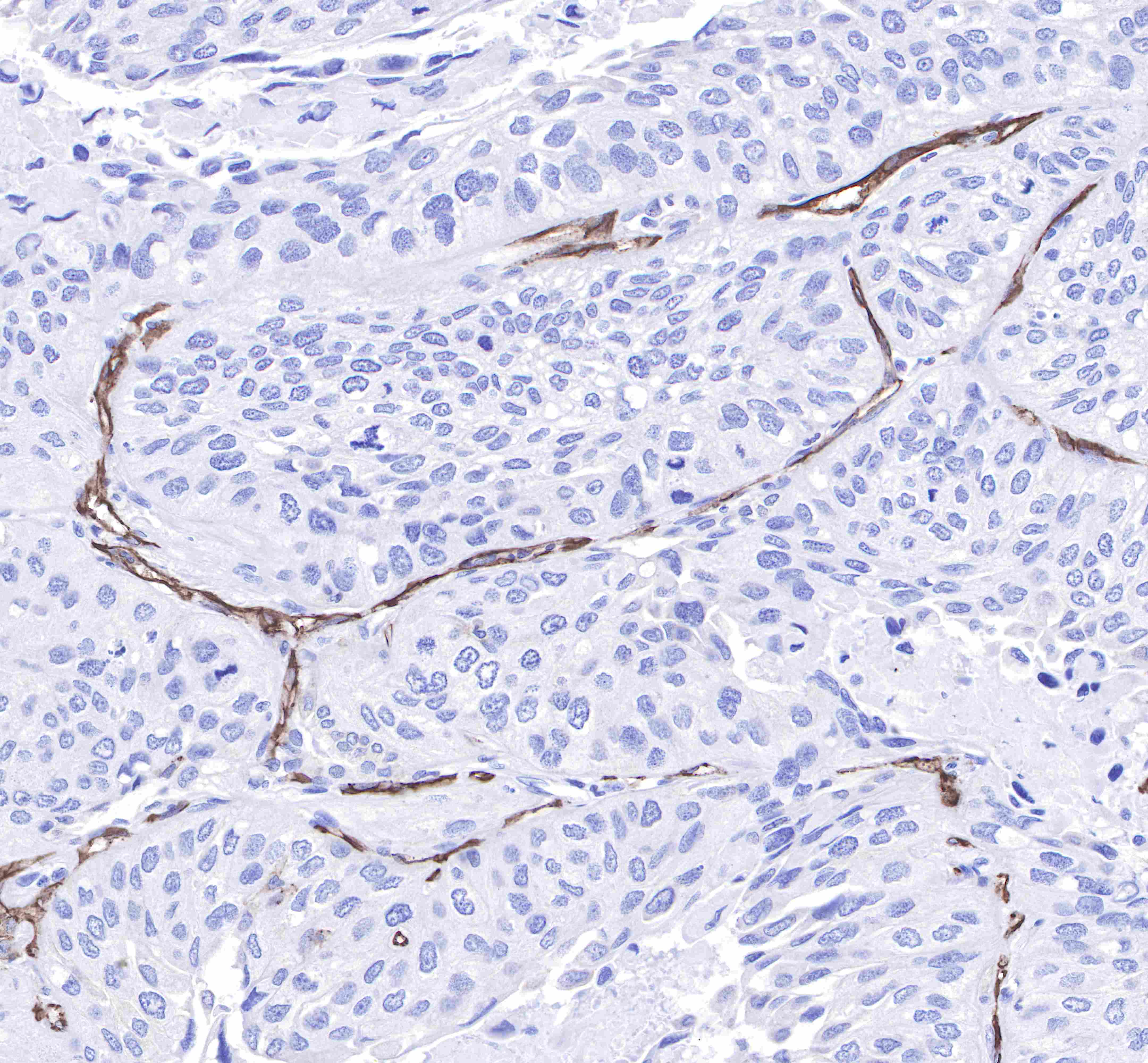
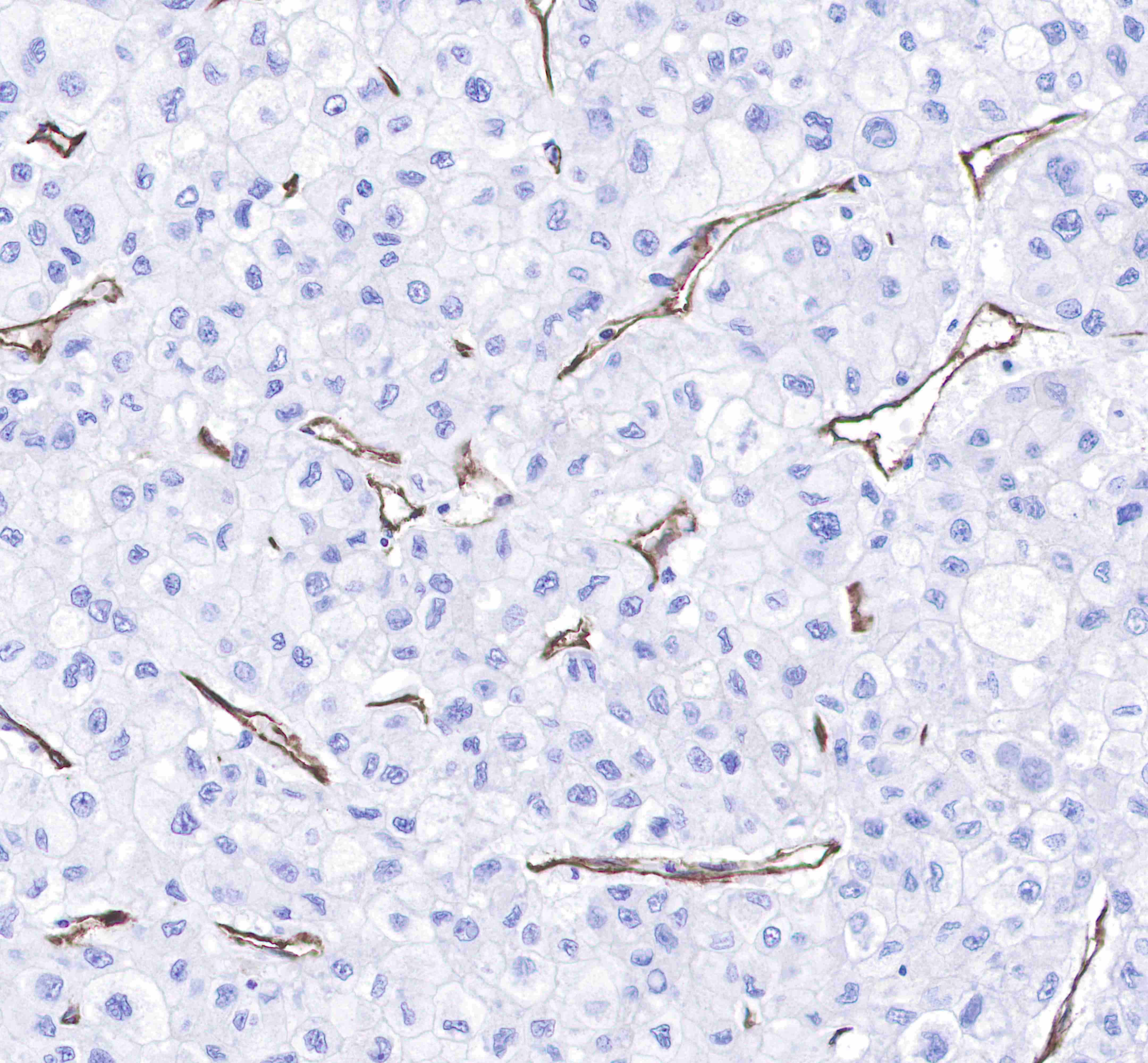
CD31 |
| Synonyms |
PECAM-1,EndoCAM, GPIIA, PECA1 |
| Immunogen |
Synthetic Peptide |
| Accession |
P16284 |
| Clone Number |
SDT-008-17 |
| Antibody Type |
Rabbit mAb |
| Isotype |
IgG |
| Application |
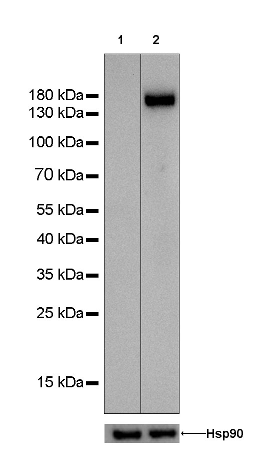
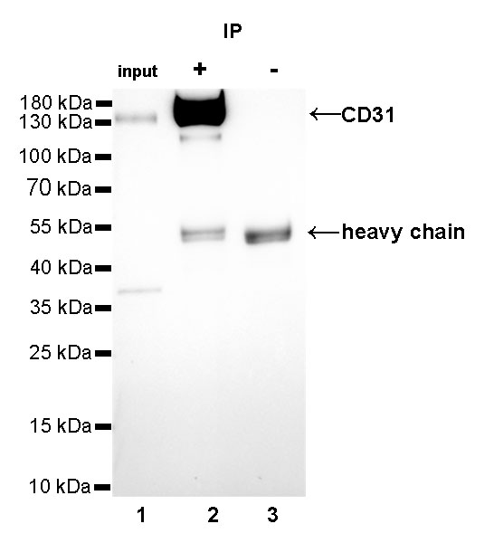
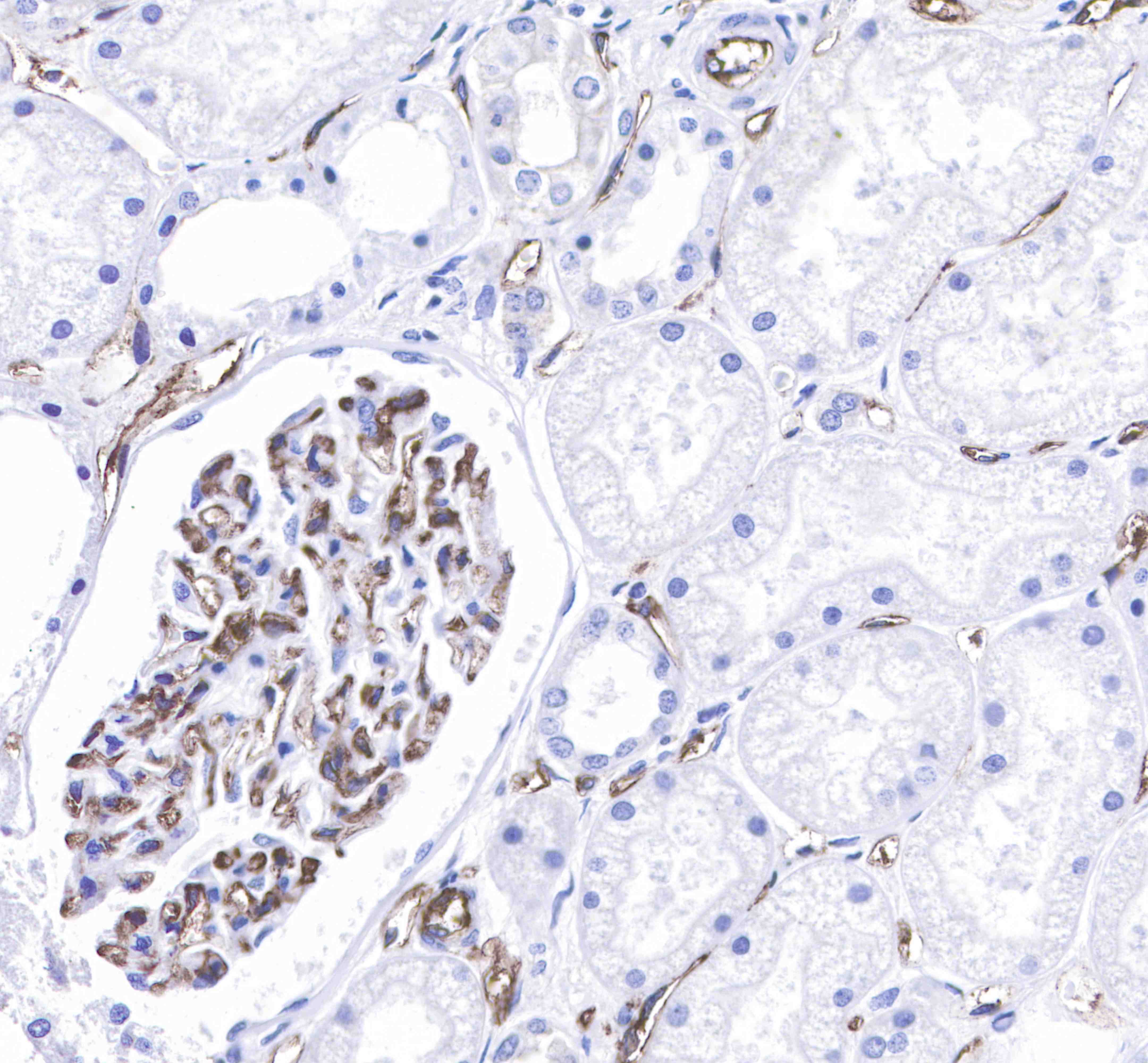
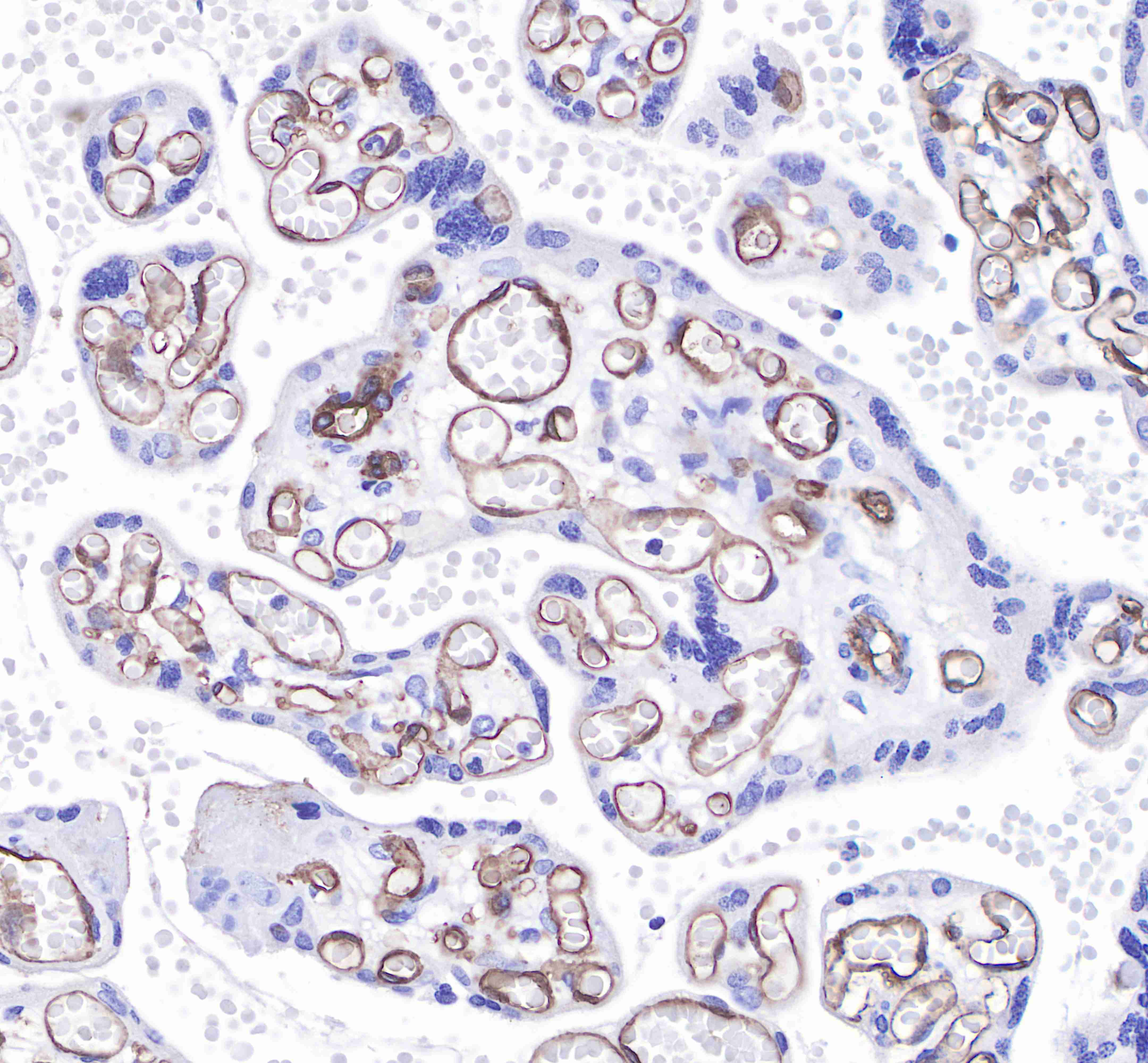
WB, IHC-P, IP |
| Reactivity |
Hu |
| Purification |
Protein A |
| Research Area |
Immunology |
| Concentration |
0.5mg/ml |
| Molecular Weight |
150kDa |
| Conjugation |
Unconjugated |
| Physical Appearance |
Liquid |
| Storage Buffer |
PBS, 40% Glycerol, 0.05%BSA, 0.03% Proclin 300 |
| Stability & Storage |
12 months from date of receipt / reconstitution, -20 °C as supplied |
Dilution
| application |
dilution |
species |
| IHC-P |
1:250 |
|
| IP |
1:25 |
|
| WB |
1:1000 |
|
Background
Cluster of differentiation 31 (CD31) also known as platelet endothelial cell adhesion molecule (PECAM-1) is a member of the immunoglobulin superfamily and is likely involved in leukocyte transmigration, angiogenesis, and integrin activation. PECAM-1 plays a key role in removing aged neutrophils from the body. PECAM-1 is found on the surface of platelets, monocytes, neutrophils, and some types of T-cells, and makes up a large portion of endothelial cell intercellular junctions. In immunohistochemistry, CD31 is used primarily to demonstrate the presence of endothelial cells in histological tissue sections. This can help to evaluate the degree of tumor angiogenesis, which can imply a rapidly growing tumor. Malignant endothelial cells also commonly retain the antigen, so that CD31 immunohistochemistry can also be used to demonstrate both angiomas and angiosarcomas. It can also be demonstrated in small lymphocytic and lymphoblastic lymphomas, although more specific markers are available for these conditions.