Product Specification
| Host |
Rabbit |
| Antigen |
Calponin-1 |
| Synonyms |
Basic calponin, Calponin H1 (smooth muscle), CNN1,CALP |
| Immunogen |
Synthetic Peptide |
| Accession |
P51911 |
| Clone Number |
SDT-080-78 |
| Antibody Type |
Rabbit mAb |
| Application |
WB, IHC-P, ICC, IF |
| Reactivity |
Hu, Ms, Rt |
| Purification |
Protein A |
| Concentration |
0.5mg/ml |
| Conjugation |
Unconjugated |
| Physical Appearance |
Liquid |
| Storage Buffer |
PBS, 40% Glycerol, 0.05%BSA, 0.03% Proclin 300 |
| Stability & Storage |
12 months from date of receipt / reconstitution, -20 °C as supplied |
Dilution
| application |
dilution |
species |
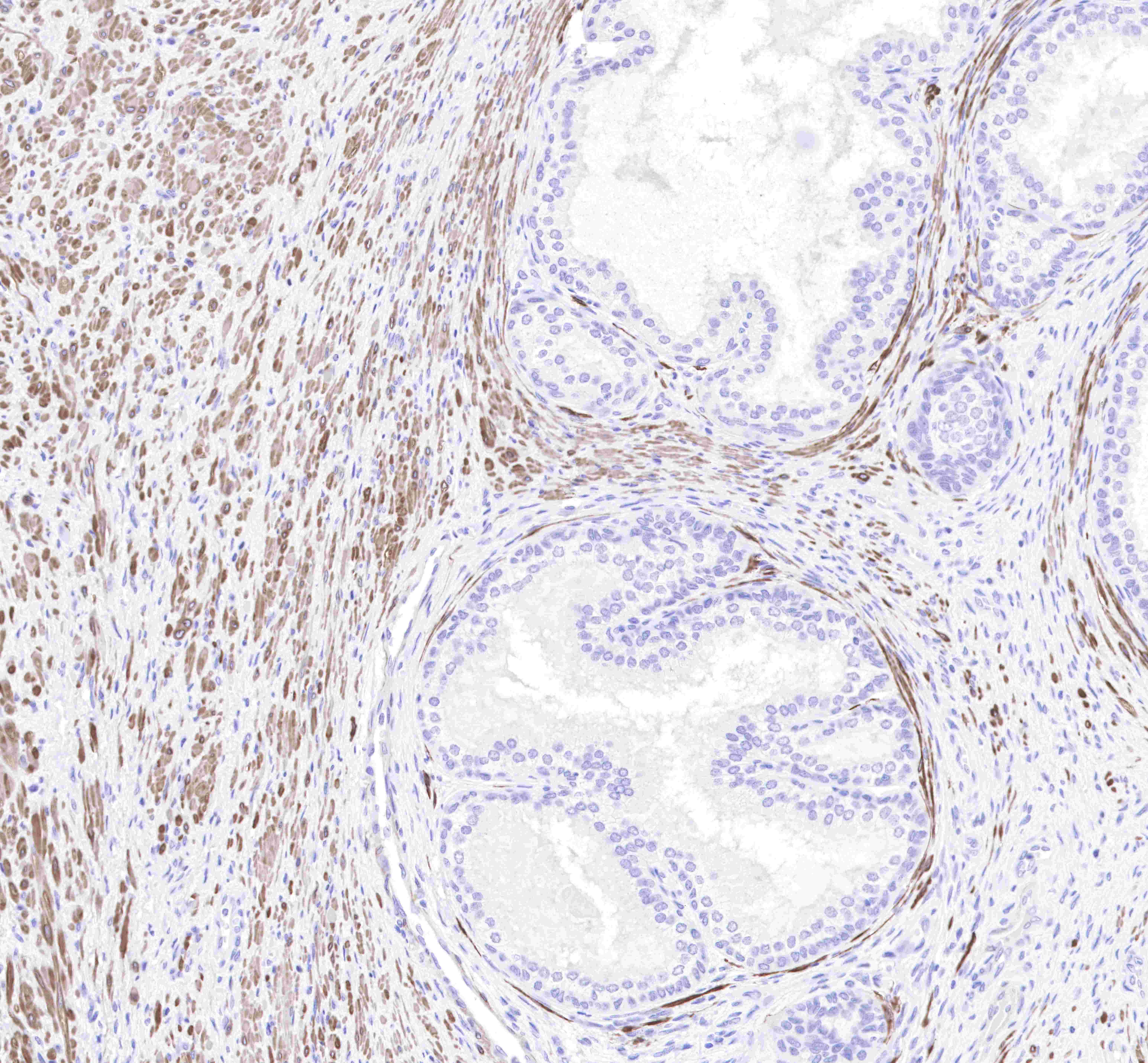
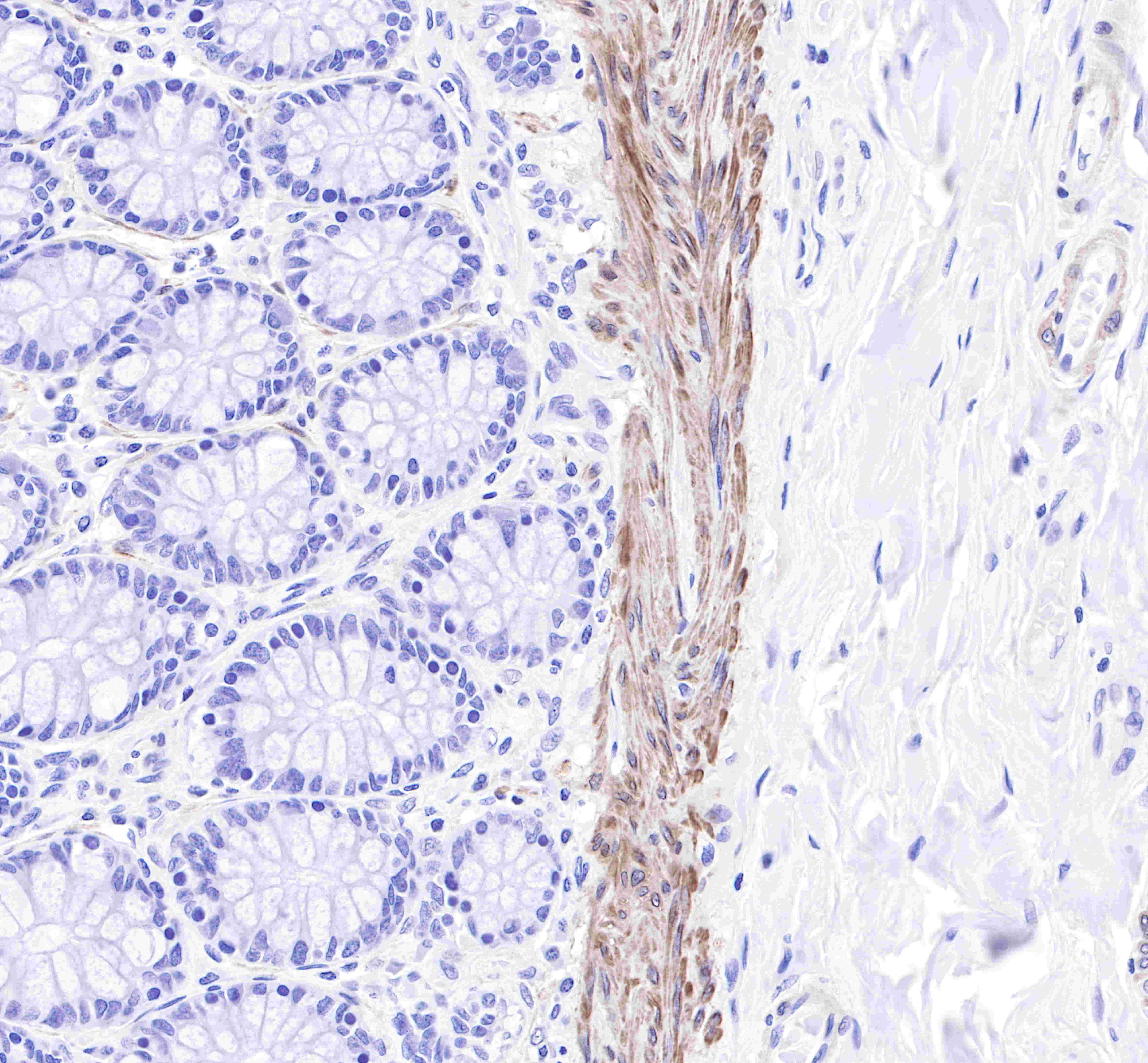
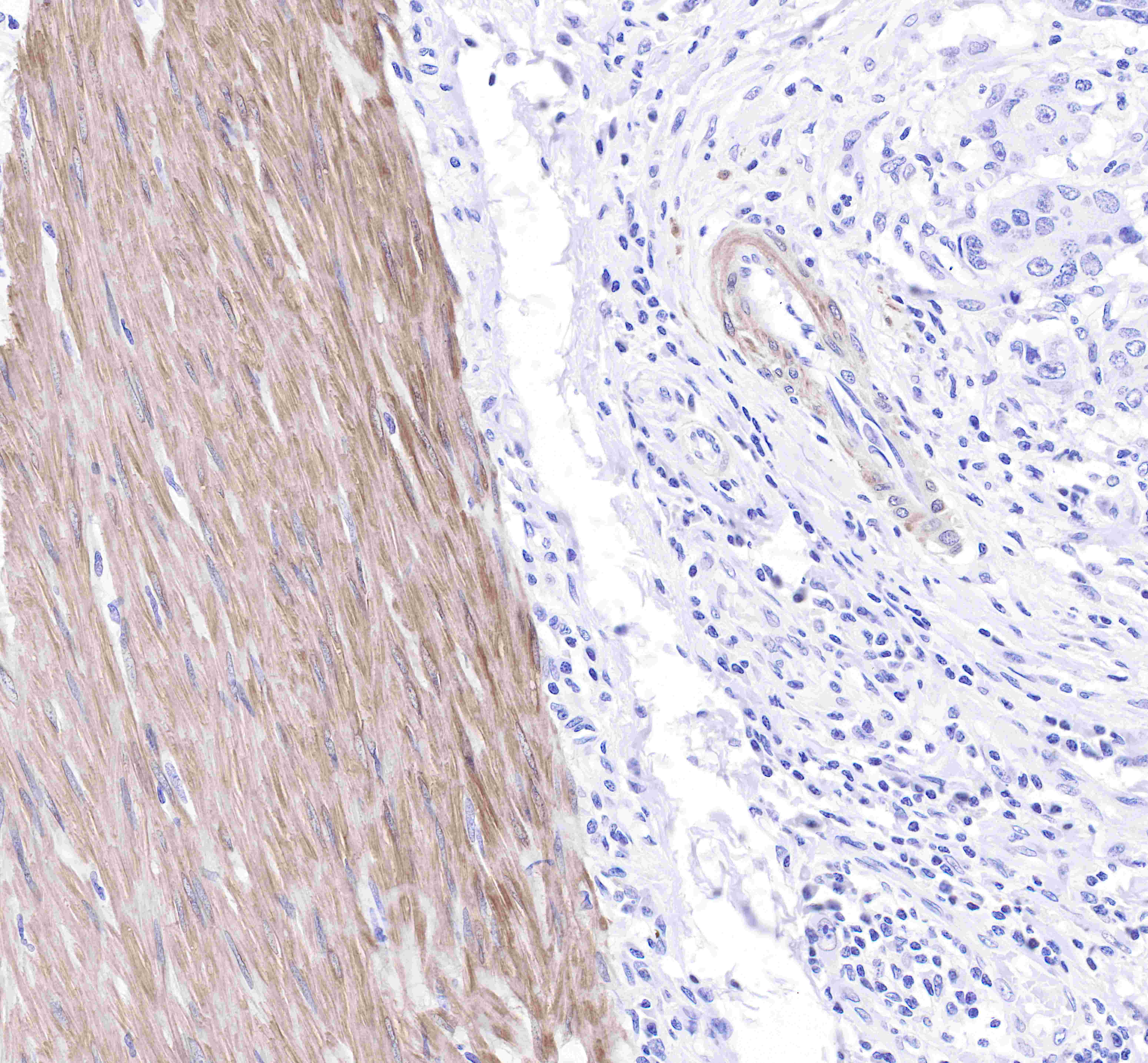
| IHC-P |
1:2000 |
|
| ICC |
1:250 |
|
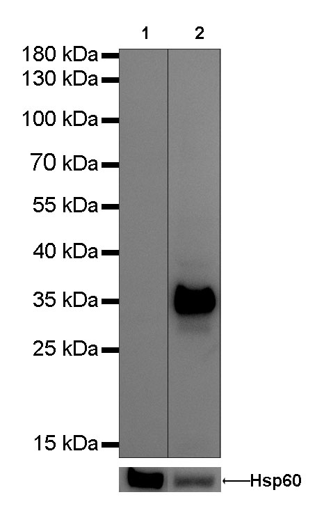
| WB |
1:1000 |
|
| IF |
1:1000 |
|
Background
Calponin-1 is a 34 kDa actin-binding protein with regions of sequence homology to cardiac troponin I and T. It is located in the cytoskeleton and contractile apparatus of differentiated smooth muscle cells. There are 3 isoforms of calponin: calponin-1 (h1 or basic), calponin-2 (h2 or neutral), and calponin-3 (h3 or acidic). Calponin-1 is implicated in the regulation of smooth muscle contraction by mediating intracellular signaling responses to some vasoconstrictors by acting as a contractile scaffold protein. This mechanism is based on results in aortic smooth muscle cells stimulated with phenylephrine, which showed calponin-1 connecting protein kinase C and ERK1/2 pathways to promote contractility.