Human TREM2 ELISA Kit
Human TREM2 ELISA Kit
Price:
Regular price
$447 USD
Regular price
Sale price
$447 USD
Unit price
per
For shipping services or bulk orders, you may request a quotation.
Secure checkout with

View full details
Product Details
Product Details
Product Specification
| protein | TREM2 | ||||||||||||||||||||||||||||||||||||||||
| Usage |
1. Self-brought consumables: 1 , plate reader (capable of measuring 450 nm Absorbance at) 2 , multi-channel pipette or automatic plate washer 3 , incubator, cryogenic centrifuge 4 Adjustable pipette gun and gun tip 5 Deionized water 2. Reagent preparation: Standard Diluent : Ready-to-use; Before use, equilibrate to room temperature; 2-8℃ Save. Standard Diluent Used to dilute standard. 1×Assay Buffer : Before use, equilibrate to room temperature and press with deionized water 1 : 10 Proportional dilution of Assay Buffer (10×) get 1×Assay Buffer , mix gently to avoid foaming. 2-8℃ Preservation, this solution can be stable 30 Heaven. 1×Assay Buffer For diluting serum and plasma samples, HRP-conjugated Human TREM2 Detect Antibody (100×) 。 Human TREM2 Standard : The label superscript added to the standard focuses on suspension volume reconstitution in deionized water Human TREM2 Standard get 20 ng/mL Standards. Before performing dilution, hold the standard at least 15 min And gently shake, after reconstitution Standard Available at -20℃ Save 1 Months, redissolved and used 1 Discard after times. 1×HRP-conjugated Human TREM2 Detect Antibody : Mix thoroughly before dilution. Depending on the amount required for standard and sample, use in clean plastic tubes 1×Assay Buffer Right HRP-conjugated Human TREM2 Detect Antibody (100×) Conduct 1:100 Dilution. 1×HRP-conjugated Human TREM2 Detect Antibody Should be after dilution 30 min Internal use. HRP Substrate (TMB) : Ready-to-use; Before use, equilibrate to room temperature; 2-8℃ Store in the dark from light. Stop Solution : Ready-to-use; Before use, equilibrate to room temperature; 2-8℃ Save. 1×Wash Buffer: Equilibrate to room temperature before use, Wash Buffer (20×) Performed with deionized water 1:20 Dilution to obtain 1×Wash Buffer 。 Mix gently to avoid blistering, store at room temperature, this solution is stable 30 Heaven. Standard curve settings: As shown in the table below, use Standard Diluent Will 20 ng/mL The standard was diluted to 10 、 5 、 2.5 、 1.25 、 0.63 、 0.31 、 0.16 And 0 ng/mL Of Human TREM2 Standards.
Note: Each experiment requires a new formulation of standards. 3. Sample preparation: 1 Cell culture supernatant: centrifuge to remove particles, immediately measure or dispense -20℃ Store and avoid repeated freezing and thawing. 2 Serum: Use a serum separation tube and allow the sample to coagulate at room temperature 30 min , then 1,000 g Centrifugation 15 min 。 Take the upper layer serum for immediate determination or dispensing -20℃ Store and avoid repeated freezing and thawing. 3 Plasma: use EDTA , heparin or citrate as an anticoagulant. After collection 30 min Inner 1,000 g Centrifugation 15 min 。 Immediate determination or dispensing -20℃ Store and avoid repeated freezing and thawing. CAUTION: Do not use specimens with severe hemolysis or lipidemia. If the sample is to be in 24 h Used within, you can store it in 2 To 8℃ , avoid repeated freezing and thawing. Frozen samples should be slowly warmed to room temperature and gently mixed prior to determination. 4. Experimental steps: 1 Remove the excess slats from the plate frame, place them back in the aluminum foil bag containing the desiccant pack, and reseal them. 2 The liquid in each well was removed and washed. Add per well using a multi-channel pipette or automatic washer 300 μL 1×Wash buffer Let stand and soak 30 s , performing washing. Complete removal of the liquid in each step is essential to obtain good performance. After the last wash, the plate was inverted and blotted dry with a clean paper towel to remove any remaining 1×Wash Buffer 。 Use the plate immediately after the plate wash is completed and do not allow the plate to dry. 3 , Add standard: add standard well 100 μL Diluted standards. 4 Add sample: serum / Plasma: sample well addition 80 μL 1×Assay Buffer And 20 μL Pre-diluted samples; Cell culture supernatant: sample well addition 100 μL Cell culture supernatant (See sample preparation for details). 5 , add detection resistance, add each well 50 μL 1×HRP-conjugated Human TREM2 Detect Antibody , guarantee step 3 、 4 、 5 Add samples continuously without interruption. The sample loading process is 15 min Within completion. Plate was coated and incubated at room temperature 2 h 。 6 Repeat steps 2 Washing in 6 Times. 7 , adding per well 100 μL HRP Substrate (TMB) 。 The plate was coated and incubated at room temperature protected from light 5-30 min 。 8 , adding per well 100 μL Stop Solution , Stop Solution Should be in accordance with the TMB The same order is added to the plate. The color in the hole should change from blue to yellow. If the color in the hole is green or the color changes unevenly, tap the plate lightly to ensure thorough mixing. 9 , in 30 min Within, measure each well 450 nm The absorbance value at. Note: 5-30 min The color development time is within the empirical range. For each specific experiment, the approximate color development time can be determined according to the following conditions. ( 1 ) Visual observation: standard song S5 The holes are light blue, Blank When the hole has no obvious blue color, it can be terminated; ( 2 ) Instrument judgment: 630 nm At left and right wavelengths, standard curve S1 Porous OD Value reaches 0.5 - 0.7 、 S5 Porous OD Value reaches 0.05 - 0.08 、 Blank Porous OD Value less than 0.05 It can be terminated. 5. Result calculation: 1 Calculate the multiwell average of each standard and sample OD Value, and subtract the zero concentration ( Std.8 ) average OD Value. 2 , Drawing of standard curve: take the standard concentration as x Axis, the average absorbance of each standard is y Axis, plot the standard curve. You can use mapping software to create standard curves. Note: If the sample is diluted, the concentration read from the standard curve must be multiplied by the dilution factor. 6. Results display: Typical standard curve ( R2≥0.99 ) 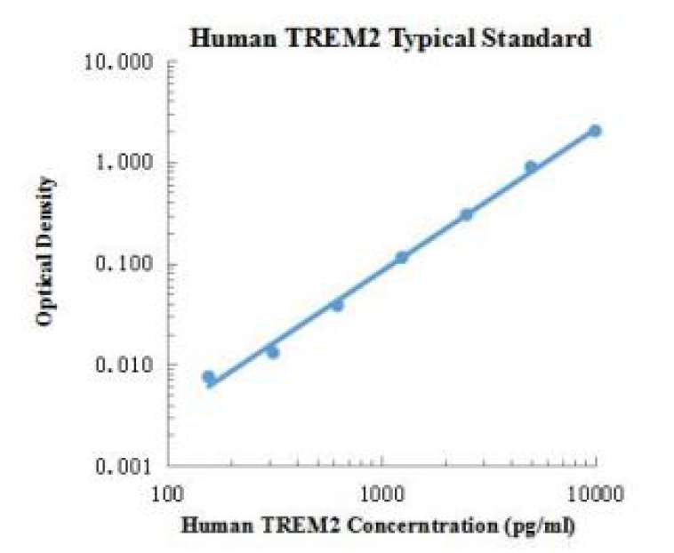 Figure1. 96 Well plate analytical Human TREM2 Standard curve. The data and curves are for reference only, and experimenters need to establish standard curves according to their own experiments. |
||||||||||||||||||||||||||||||||||||||||
| Sensitivity | 0.16 ng/mL | ||||||||||||||||||||||||||||||||||||||||
| Species Reactivity | Human | ||||||||||||||||||||||||||||||||||||||||
| Source | Human | ||||||||||||||||||||||||||||||||||||||||
| Synonym | Human TREM-2 ELISA Kit | ||||||||||||||||||||||||||||||||||||||||
| Detection Type | Sandwich method | ||||||||||||||||||||||||||||||||||||||||
| Description | Myeloid cell trigger receptor 2 (TREM2) is a single Ig domain receptor that is expressed in macrophages and dendritic cells, but not in granulocytes or monocytes. It is most abundantly expressed in the basal ganglia, corpus callosum, medulla oblongata and spinal cord, while microglia in the central nervous system are the most dominant cell type producing TREM2. TREM2 plays a role in chronic inflammation and can stimulate the production of constituent rather than inflammatory chemokines and cytokines. TREM2 forms a receptor signaling complex with TYROBP to activate the immune response of macrophages and dendritic cells. It is also associated with the adaptor protein DAP12 to transmit activation signals, which play a role in innate and adaptive immune responses. TREM2 Signaling is also an important way to promote colonic wound healing. TREM2 mutations increase the risk of neurodegenerative diseases such as Alzheimer's disease, amyotrophic lateral sclerosis, and Parkinson's disease. The Human Myeloid Cell Trigger Receptor 2 ELISA Quantification Kit employs a double antibody sandwich method to quantify human myeloid cell trigger receptor 2 in a sample. The plate is coated with an antibody specific to human myeloid cell trigger receptor 2, and the sample or standard and the specific detection antibody labeled with HRP are added to the wells of the plate. The human myeloid cell trigger receptor 2 binds to the solid phase antibody and the detection antibody coated on the plate. After washing, a chromogenic substrate (TMB) was added, and the TMB became blue under HRP catalysis and turned yellow after addition of the stop solution. The OD value was measured with a microplate reader at 450 nm wavelength, and the concentration of human myeloid cell trigger receptor 2 was directly proportional to the OD450 value. |
||||||||||||||||||||||||||||||||||||||||
| Composition |
|
||||||||||||||||||||||||||||||||||||||||
| General Notes | 1. If Standard Diluent and Assay Buffer (10 ×) turn yellow in color and have a small amount of precipitation, it is because the reagent contains serum. Just centrifuge to remove the precipitation, which will not affect normal use. 2. Do not mix components with different batch numbers and different manufacturers; Failure to do so may lead to abnormal results. 3. Change the tip frequently to avoid cross-contamination between the components. 4. To ensure accurate results, the plate must be properly adhered during the incubation step. 5. Stop Solution is corrosive to a certain extent, so please take protective measures when operating. |
||||||||||||||||||||||||||||||||||||||||
| Instructions | Serum (plasma), cell culture supernatant | ||||||||||||||||||||||||||||||||||||||||
| Storage Temp. | 2-8 ℃, protected from light, valid for 24 months. | ||||||||||||||||||||||||||||||||||||||||
| Test Range | 0.16 ng/mL-10 ng/mL |