Human PD-1 ELISA Kit
Human PD-1 ELISA Kit
Price:
Regular price
$450 USD
Regular price
Sale price
$450 USD
Unit price
per
For shipping services or bulk orders, you may request a quotation.
Secure checkout with

View full details
Product Details
Product Details
Product Specification
| protein | PD-1 | ||||||||||||||||||||||||||||||||||||||||||||||||||||||||||||||||||||
| Usage |
1. You need to bring your own test equipment 1. Microplate reader (measurable 450nm Absorption value of detection wavelength and 540nm Or 570nm Absorption value of corrected wavelength) 2. High precision liquid dispenser and disposable tip 3. Distilled or deionized water 4. Bottle washer (spray bottle), multi-channel plate washer or automatic plate washer 5. 500mL Measuring cylinder 2. Preparation before the experiment 1 Sample collection and storage Cell culture supernatant: Particulate matter should be removed by centrifugation; Test the sample immediately. If the sample is not tested in time after collection, it is recommended to pack it according to the amount used once and store it frozen in -20℃ In the refrigerator, avoid repeated freezing and thawing. The sample may need to be used with a diluent (1×) Dilution. Serum: Using serum separation tubes (SST) Collect samples and place samples at room temperature 30 Minutes. Centrifugation 15 Minutes, with a rotation speed of 1000 g。 The serum was removed immediately and tested immediately. If the sample is not tested in time after collection, it is recommended to pack it according to the amount used once and store it frozen in ≤-20℃ In the refrigerator, avoid repeated freezing and thawing. The sample may need to be used with a diluent (1×) dilution. Plasma: Using EDTA, heparin or citric acid as an anticoagulant to collect plasma, after collection 30 Centrifuge within minutes 15 Minutes, with a rotation speed of 1000 g, and detect it immediately. If the sample is not tested in time after collection, it is recommended to pack it according to the amount used once and store it frozen in ≤-20℃ In the refrigerator, avoid repeated freezing and thawing. The sample may need to be used with a diluent (1×) dilution. 2 Preparation work before testing Please place all reagents and samples at room temperature before use and let them stand 15 Minutes. It is recommended that all experimental samples and standards be repeatedly tested 1× Wash solution preparation: The concentrated wash solution in the kit is 20× Mother liquor should be diluted with distilled water before use to 1× Working fluid. Example: Take 10ml Concentrated wash +190mL Distilled water to volume to 200mL In actual operation, the usage amount can be calculated first, and then prepared. 1× Buffer preparation for dilution: The concentration and dilution buffer in the kit is 10× Mother liquor, dilute with distilled water before use to 1× Working fluid. Example: Take 3ml Buffer for concentration and dilution +27mL Distilled water to volume to 30mL。 In actual operation, the required amount of dilution buffer solution can be calculated according to the sample dilution factor, and then prepared. Antibody detected: Centrifuge the dry powder to the bottom of the tube and use 110μL Buffer for dilution (1×) Dissolve and let stand at room temperature 5min Later obtained 100× Mother liquor; Before use, dilute to 1× Working fluid. According to the amount per well 100μL Calculate the required volume. Example: Used 10 Hole, then take 10μL Of 100 Double working concentration of detection antibody, using dilution buffer (1×) Constant volume to 1mL, obtained 1ml Of 1× Detection antibody at working concentration SA-HRP: SA-HRP For 40× Mother liquor, use dilution buffer before use (1×) Diluted and formulated 1× Working fluid, the required amount per hole is 100 μL。 Example: Used 10 Hole, then take 25μL Of 40× Mother liquor +975uL Buffer for dilution (1×) Constant volume to 1mL, obtained 1ml Of 1× Detection antibody at working concentration. Color development solution: Per hole 100 μL Calculate the dosage required for the current test, take out the corresponding volume of color developer, and protect it from light; The developer removed is for the same day use only. Standard: Dilution buffer for lyophilized standards (1×) re-dissolve, Re-dissolve Volume 1000μL, obtaining a concentration of 10000pg/mL Standard mother liquor. Gently shake at least 5 Minutes, it is fully dissolved. Add to each dilution tube 300 μL Dilution Buffer (1×)。 Make serial dilutions of the standard mother liquor according to the figure below, and each tube must be thoroughly mixed before pipetting to the next tube. Standard mother liquor without dilution can be used as the highest point of standard curve (10000pg/mL), Dilution Buffer (1×) Can be used as a standard curve zero (0 pg/mL)。 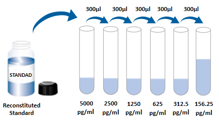 3. Experimental operation steps 1 Prepare all required reagents and standards; 2 Take out the microplate from the sealed bag that has been balanced to room temperature. Please put the unused slats back into the aluminum foil bag and re-seal; 3 Adding to the microplate 300μL Washing liquid, let stand and soak 30 Seconds, discard the lotion and pat the microplate dry on absorbent paper. Please use it immediately and do not let the microplate dry; 4 Add different concentration standard substances, experimental samples or quality control substances into corresponding wells, and each well 100μL。 Seal the reaction wells with plate sealing tape and incubate at room temperature 2 hour; 5 Suck off the liquid in the plate, and use a bottle washing machine, a multi-channel plate washer or an automatic plate washer to wash the plate. Washing solution per well 300μL Then the wash liquid in the plate is aspirated off. Repeat Operation 3 Times. Trying to absorb the residual liquid as much as possible every time you wash the plate will help to get good experimental results. At the end of the last plate washing, please suck all the liquid in the plate or turn the plate upside down, and pat all the residual liquid dry on absorbent paper; 6 Adding in each microwell 100μL Detect antibodies. Seal the reaction wells with plate sealing tape and incubate at room temperature 2 Hour; 7 Repeat the first 5 Step washing operation; 8 Adding in each microwell 100μL SA-HRP, room temperature incubation 20 Minutes. Be careful to avoid light; 9 Repeat the first 5 Step washing operation; 10 Adding in each microwell 100 μL Chromogenic solution, incubate at room temperature 5-30 Minutes, pay attention to avoiding light; 11 Adding in each microwell 50μL Stop solution, the color of the solution in the well will change from blue to yellow. If the color of the solution changes to green or the color changes are inconsistent, pat the microplate gently to mix the solution evenly; 12 After adding the stop solution 30 Within minutes, measured using a plate reader 450nm Absorbance value, set 540nm Or 570nm As a correction wavelength. If dual-wavelength correction is not used, the accuracy of the results may be affected; 13 Calculation results: add the corrected absorbance values of each standard and sample (OD450-OD540/OD570), average of repeated well readings and then subtract the average zero standard OD Value. Using computer software for four-parameter logic (4-PL) Curve fitting creates a standard curve. Another way is to plot the standard concentration and make the logarithm with the corresponding OD The values were logarithmic to generate a curve, and the best fit line was determined by regression analysis. This process can generate a data fit that is sufficiently useful but less accurate. If the sample is diluted, the concentration should be calculated by multiplying the dilution factor. 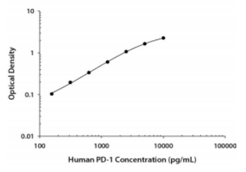 4. Kit parameters 1 Recovery rate Spiked different levels of human in cell culture medium samples PD-1 The recovery rate was determined. The recoveries range from 85.0-111.9%, the average recovery was in 100.9%。 Inspired human serum samples with different levels of human PD-1 The recovery rate was determined. The recoveries range from 99.3-121.1%, the average recovery was in 111.0%。 2 Sensitivity Person PD-1 The lowest measurable dose (MDD) Generally less than 18.9 pg/mL。 The lowest measurable value is determined according to 20 The corresponding concentration is calculated by adding two standard deviations to the mean value of the zero-point absorbance values of each standard curve. 3 Correction This ELISA The kit NS0 Expressed high purity recombinant human PD-1 Corrected by protein. 4 Linearity 4 Different samples were spiked with high concentrations of human PD-1, Then using a diluent (1×) Dilute the sample to the detection range, Its linearity was determined.
5 Specificity, This ELISA Method can detect natural and recombinant human PD-1 Egg whites. The following factors were mixed with diluent (1×) Formulated into 100 ng/mL Concentration to detect human PD-1 Cross-reactivity of. Will 100 ng/mL Interference factors incorporated into the intermediate range of recombinant human PD-1 In the reference substance, To detect human PD-1 Of interference. No significant cross-reactivity or interference was observed.
5. Analysis of frequently asked questions 1 Whiteboard (after color rendering is completed, no color appears)
2, flower plate (blank, negative positive control normal, but specimen well OD Values are significantly higher)
5. Experimental flow chart 
|
||||||||||||||||||||||||||||||||||||||||||||||||||||||||||||||||||||
| Species Reactivity | Human | ||||||||||||||||||||||||||||||||||||||||||||||||||||||||||||||||||||
| Theory | This kit adopts double antibody sandwich enzyme-linked immunosorbent detection technology. Specific anti-human PD-1 antibodies were pre-coated on high affinity plates. The standard substance, the sample to be tested and the biotinylated detection antibody are added to the wells of the enzyme label plate. After incubation, PD-1 present in the sample binds to the solid phase antibody and the detection antibody to form an immune complex. After washing to remove unbound material, horseradish peroxidase-labeled Streptavidin-HRP was added. After washing, a chromogenic substrate is added to protect the color from light. A stop solution was added to stop the reaction, and the absorbance value was measured at a wavelength of 450 nm (reference calibration wavelength of 540 nm or 570 nm). | ||||||||||||||||||||||||||||||||||||||||||||||||||||||||||||||||||||
| Source | Human | ||||||||||||||||||||||||||||||||||||||||||||||||||||||||||||||||||||
| Wells | 0 | ||||||||||||||||||||||||||||||||||||||||||||||||||||||||||||||||||||
| Composition |
|
||||||||||||||||||||||||||||||||||||||||||||||||||||||||||||||||||||
| Background | Programmed death-1 receptor (PD-1), also known as CD279, is a type I transmembrane protein of the CD28 family of immunomodulatory receptors, other members of which include CD28, CTLA-4, ICOS, and BTLA. Its cytoplasmic tail contains two tyrosine residues that make up the tyrosine-based immunoreceptor inhibitory domain (ITIM) and switch domain (ITSM), which are important for mediating PD-1 signal transduction. As a monomeric receptor, PD-1 can interact with its ligands PD-L1 (B7-H1) and PD-L2 (B7-DC) in a ratio of 1: 1. PD-1 is expressed in activated T cells, B cells, monocytes and dendritic cells, while PD-L1 is expressed in non-hematopoietic cells (such as lung endothelial cells and hepatocytes) in addition to the above cells. The combination of PD-L1 and PD-1 can induce co-inhibitory signals, promoting T cell inactivation, apoptosis, and functional failure. Therefore, PD-1 and PD-L1 interaction is a key regulator of immune response threshold and peripheral immune tolerance. Blocking the interaction of PD-1 and PD-L1 through antibody or gene regulation can accelerate tumor eradication, suggesting the potential of PD-1 as a target for cancer immunotherapy. | ||||||||||||||||||||||||||||||||||||||||||||||||||||||||||||||||||||
| General Notes | 1. Only for scientific research, not for in vitro diagnosis; 2. Please use it within the validity period of the kit; 3. The components of different kits and kits with different batch numbers cannot be mixed; 4. If the sample value is greater than the highest value of the standard curve, the sample should be diluted with diluent (1 ×) and re-tested; If the cell culture supernatant sample needs to be distributed and diluted, cell culture medium can be used for other intermediate dilutions except for the last step of dilution with diluent; 5. Differences in test results can be caused by a variety of factors, including the operation of the experimenter, the use of the pipette, the plate washing technique, the reaction time or temperature, the storage of the kit, etc. 6. The terminating solution in the kit is an acidic solution. Please protect your glasses, hands, face and clothes when using it.
|
||||||||||||||||||||||||||||||||||||||||||||||||||||||||||||||||||||
| Storage Temp. | The unopened kit is stored at 2-8 ℃ and has a validity period of one year. |
Picture
Picture
Immunohistochemistry