Cytotoxic drug activity assay: Hela cells were seeded at 5,000 cells per well in a clear-bottom 96-well plate and treated with different concentrations of docetaxel. After 65 hours, the plates were removed, and sequential fluorescence-based cell viability assays—CTG cell viability assays—were performed using volumes of 60 μL medium, 60 μL fluorescence reagent, and 60 μL CTG reagent. The readings from the untreated control wells were set to 100%, the percent reading for each drug-treated well was calculated, and a graph was generated to determine the IC50.
A. and B. represent the fluorescence detection results for a foreign brand's comparable product and UA CTF, respectively, while C. and D. are the CTG detection results obtained after CTF testing of A. and B., respectively. Using two different kits and two distinct detection methods, the IC50 values were consistently very close, ranging from 6 to 9 nM. These results indicate that the fluorescent cell viability assay reagent is compatible with the CTG assay reagent, and that the CTF assay reagent exhibits performance comparable to that of its foreign counterpart.
UA-Glo® Fluorescent Cell Viability Assay
UA-Glo® Fluorescent Cell Viability Assay

Product Details
Product Details
Product Specification
| Synonyms | 荧光细胞活力检测试剂盒 |
| Stability & Storage | Store at -20℃ or below. Valid for 12 months. |
Background
There are multiple methods for cell viability assays, such as dye exclusion, ATP concentration changes, and enzyme activity measurements. Cell assays sometimes require mechanistic studies of cytotoxicity and/or the inclusion of internal controls, leading to the need for multiplex analysis—where multiple assay methods are applied to the same sample. These methods should have distinct detection mechanisms and not interfere with each other, enabling comparative analysis to reveal cytotoxicity mechanisms or eliminate systemic variations. Our UA-Glo® Fluorescent Cell Viability Assay detects cell viability based on the cleavage of a peptide-fluorophore substrate by proteases exclusively present in live cells, generating a fluorescent signal. When cell membrane integrity is compromised, the protease loses activity, and no fluorescence is produced. The UA-Glo® Fluorescent Cell Viability Assay operates on a different principle than our UA-Glo® Luminescent Cell Viability Assay (Catalog No. UA070103), allowing it to detect earlier and more subtle cellular damage. The two assays are compatible for multiplex detection. Additionally, the UA-Glo® Fluorescent Cell Viability Assay is compatible with our UA-Glo® Caspases 3/7 Apoptosis Assay Kit (Catalog No. UA079012), enabling multiplex detection to determine the mechanisms of cytotoxicity.
Components
Protease substrate and buffer mixture are dispensed into 10 ml or 100 ml brown bottles and 2 ml tubes, with specifications as follows:
Specification |
CTF Substrate |
CTF Buffer |
Detectable 96-well plate wells |
Detectable 384-well plate wells |
10 ml |
100μl |
10ml |
100 wells |
500 wells |
5X10ml |
5x100μl |
5x10ml |
500 wells |
2,500 wells |
2X50 ml |
2x500μl |
2x50ml |
1,000 wells |
5,000 wells |
Protocol
1. Cell Preparation
1) Seed the cells to be tested at an appropriate density in 96-well or 384-well cell culture plates. Black-walled clear-bottom plates are recommended.
2) Add the test compounds at appropriate concentrations to the wells. Maintain the concentration of organic solvents in the culture medium below 1%. Continue incubation for the required duration based on experimental needs.
2. Cell Viability Assay
1) Take out the fluorescent cell viability assay reagent. After complete dissolution in CTF buffer, equilibrate it to room temperature. Vortex the CTF substrate to ensure thorough mixing, then centrifuge briefly to collect it at the bottom of the tube.
2) Prepare the CTF reaction solution: The CTF substrate is 100x. Dilute it to 1x reaction solution with CTF buffer according to the required volume and mix well.
3) Take out the cell culture plate and add an equal volume of CTF reaction solution to each well (e.g., add 100μl of 1x CTF reaction solution to 100μl of culture medium).
4) Shake the plate gently for 20 seconds to mix, then incubate in the dark at 37°C for 60 minutes before fluorescence detection. The optimal reading time varies depending on the cell type due to differences in CTF substrate cleavage rates. The earliest reading can be taken 30 minutes after adding the reagent, but results with higher signal-to-noise ratios are typically obtained after 60 minutes or longer. The maximum recommended reading time should not exceed 3 hours.
5) Measure the fluorescence signal using a plate reader with excitation at Ex 380-400nm and emission at Em 505nm.
6) If needed, proceed with downstream multiplex assays, such as Caspases 3/7 apoptosis detection.
Guidelines
1. After initial use, it is recommended to aliquot the substrate and store it at -20°C. Prepare the reaction solution freshly each time.
2. Unless strictly validated, it is not recommended to arbitrarily change the amount of reaction reagents.
Picture
Picture
Bioactivity
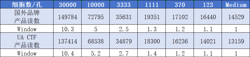
HEK293 cell counting was performed, and the cells were seeded into a 96-well plate at the cell densities indicated in the table. Fluorescent cell viability assays were conducted using UA CTF and a comparable foreign brand product, following the respective manufacturer's instructions. The values shown in the figure represent the average of two replicates measured over 60 minutes. The results indicate that the sensitivity of the two assay kits is similar.