WB result of p57 Rabbit mAb
Primary antibody: p57 Rabbit mAb at 1/1000 dilution
Lane 1: HeLa whole cell lysate 20 µg
Lane 2: HeLa treated with Dexamethasone (50nM, 16hr) whole cell lysate 20 µg
Secondary antibody: Goat Anti-Rabbit IgG, (H+L), HRP conjugated at 1/10000 dilution
Predicted MW: 57 kDa
Observed MW: 52 kDa
(This blot was developed with high sensitivity substrate)
p57 Recombinant Rabbit mAb (SDT-191-79)
p57 Recombinant Rabbit mAb (SDT-191-79)
Price:
Regular price
$100 USD
Regular price
Sale price
$100 USD
Unit price
per
For shipping services or bulk orders, you may request a quotation.
Secure checkout with

View full details
Product Details
Product Details
Product Specification
| Host | Rabbit |
| Antigen | p57 |
| Synonyms | Cyclin-dependent kinase inhibitor 1C, Cyclin-dependent kinase inhibitor p57, p57Kip2, CDKN1C, KIP2, P57-KIP2 |
| Immunogen | Synthetic Peptide |
| Location | Nucleus |
| Accession | P49918 |
| Clone Number | SDT-191-79 |
| Antibody Type | Recombinant mAb |
| Isotype | IgG |
| Application | WB, IHC-P, ICC, ICFCM, IP |
| Reactivity | Hu |
| Purification | Protein A |
| Concentration | 0.5 mg/ml |
| Conjugation | Unconjugated |
| Physical Appearance | Liquid |
| Storage Buffer | PBS, 40% Glycerol, 0.05%BSA, 0.03% Proclin 300 |
| Stability & Storage | 12 months from date of receipt / reconstitution, -20 °C as supplied |
Dilution
| application | dilution | species |
| WB | 1:1000 | |
| IHC | 1:200 | |
| ICFCM | 1:500 | |
| ICC | 1:500 | |
| IP | 1:50 |
Background
p57 is a cyclin dependent kinase inhibitor protein. Normal p57 expression (strong nuclear staining in villous cytotrophoblasts, intermediate trophoblasts and villous stromal cells) is seen in all gestations that contain maternal genetic material. p57 expression is lost in complete hydatidiform mole, while it is retained in partial hydatidiform moles and nonmolar specimens.
Picture
Picture
Western Blot
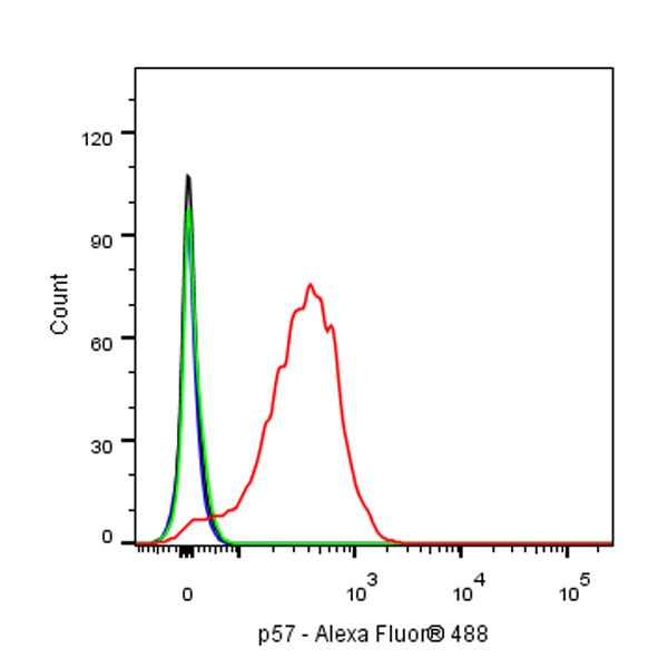
FC
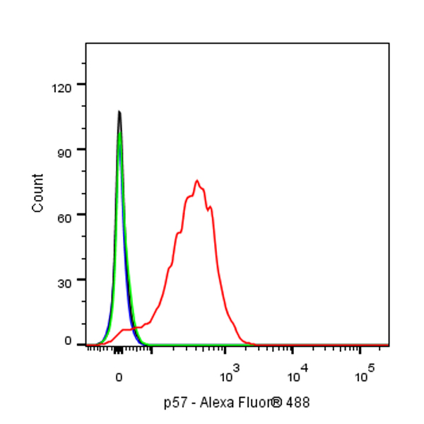
Flow cytometric analysis of 4% PFA fixed 90% methanol permeabilized HeLa (Human cervix adenocarcinoma epithelial cell) cells, treated with 50nM Dexamethasone for 16h (Red) or untreated (Green), labeling p57 at 1/500 dilution (0.1 μg) compared with a rabbit monoclonal IgG isotype control (Black) and an unlabeled control (cells without incubation with primary antibody and secondary antibody) (Blue). Goat Anti - Rabbit IgG Alexa Fluor® 488 was used as the secondary antibody.
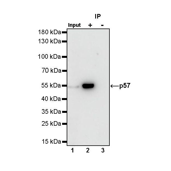
IP
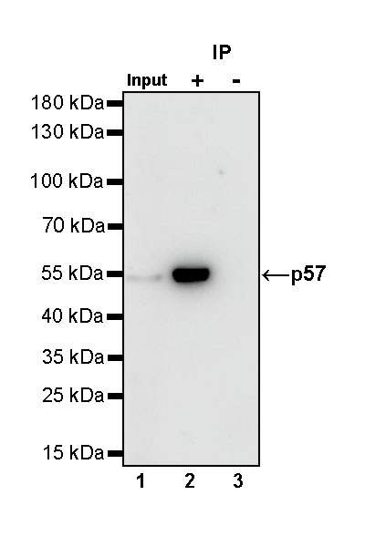
p57 Rabbit mAb at 1/50 dilution (1 µg) immunoprecipitating p57 in 0.4 mg HeLa treated with Dexamethasone (50nM, 16hr) whole cell lysate.
Western blot was performed on the immunoprecipitate using p57 Rabbit mAb at 1/1000 dilution.
Secondary antibody (HRP) for IP was used at 1/400 dilution.
Lane 1: HeLa treated with Dexamethasone (50nM, 16hr) whole cell lysate 10 µg (Input)
Lane 2: p57 Rabbit mAb IP in HeLa treated with Dexamethasone (50nM, 16hr) whole cell lysate
Lane 3: Rabbit monoclonal IgG IP inHeLa treated with Dexamethasone (50nM, 16hr) whole cell lysate
Predicted MW: 57 kDa
Observed MW: 52 kDa
(This blot was developed with high sensitivity substrate)
Immunohistochemistry
IHC shows positive staining in paraffin-embedded human placenta. Anti-p57 antibody was used at 1/200 dilution, followed by a HRP Polymer for Mouse & Rabbit IgG (ready to use). Counterstained with hematoxylin. Heat mediated antigen retrieval with Tris/EDTA buffer pH9.0 was performed before commencing with IHC staining protocol.
IHC shows positive staining in paraffin-embedded human placenta. Anti-p57 antibody was used at 1/200 dilution, followed by a HRP Polymer for Mouse & Rabbit IgG (ready to use). Counterstained with hematoxylin. Heat mediated antigen retrieval with Tris/EDTA buffer pH9.0 was performed before commencing with IHC staining protocol.
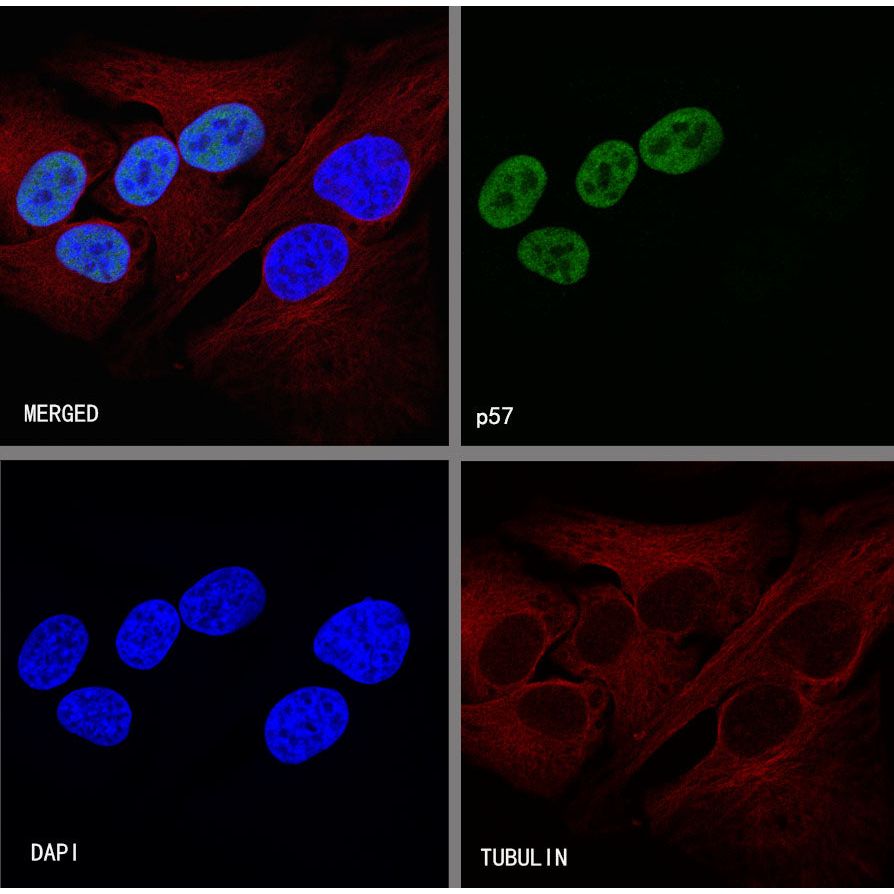
Immunocytochemistry
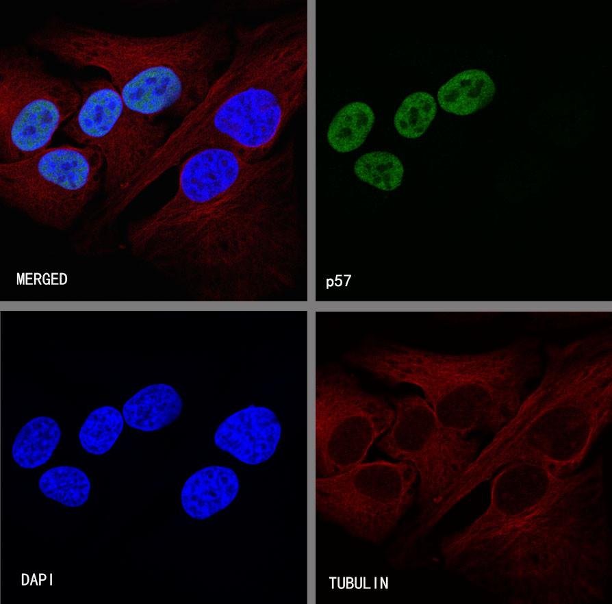
ICC shows positive staining in HeLa cells, treated with Dexamethasone(50nM, 16hr) . Anti-p57 antibody was used at 1/500 dilution (Green) and incubated overnight at 4°C. Goat polyclonal Antibody to Rabbit IgG - H&L (Alexa Fluor® 488) was used as secondary antibody at 1/1000 dilution. The cells were fixed with 4%PFA and permeabilized with 0.1% PBS-Triton X-100. Nuclei were counterstained with DAPI (Blue). Counterstain with tubulin (Red).
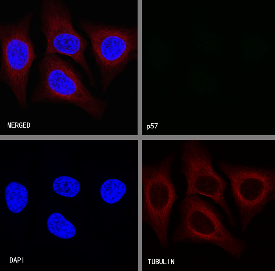
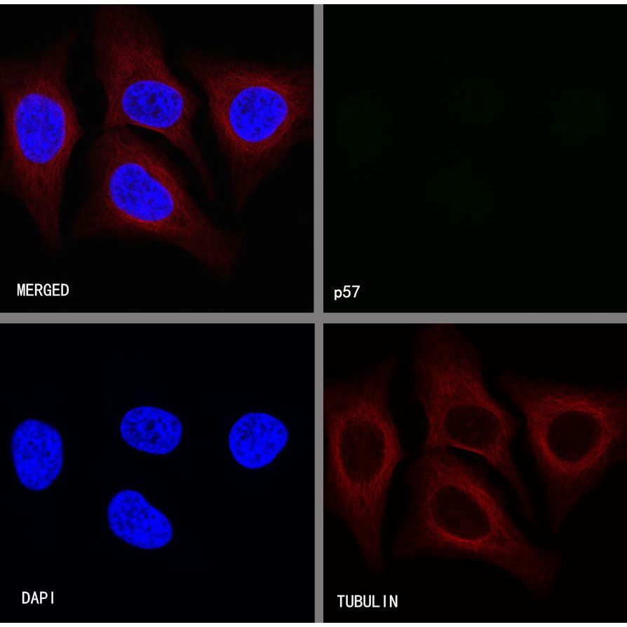
Negative control:ICC shows negative staining in HeLa cells, untreated with Dexamethasone(50nM, 16hr) . Anti-p57 antibody was used at 1/500 dilution and incubated overnight at 4°C. Goat polyclonal Antibody to Rabbit IgG - H&L (Alexa Fluor® 488) was used as secondary antibody at 1/1000 dilution. The cells were fixed with 4%PFA and permeabilized with 0.1% PBS-Triton X-100. Nuclei were counterstained with DAPI (Blue). Counterstain with tubulin (Red).