WB result of XBP-1s Recombinant Rabbit mAb
Primary antibody: XBP-1s Recombinant Rabbit mAb at 1/1000 dilution
Lane 1: untreated HeLa whole cell lysate 20 µg
Lane 2: HeLa treated with 2 μg/ml Tunicamycin for 8 hours whole cell lysate 20 µg
Secondary antibody: Goat Anti-rabbit IgG, (H+L), HRP conjugated at 1/10000 dilution
Predicted MW: 29 kDa
Observed MW: 60 kDa
This blot was developed with high sensitivity substrate
XBP-1s Recombinant Rabbit mAb (S-2341-53)
XBP-1s Recombinant Rabbit mAb (S-2341-53)
Price:
Regular price
$100 USD
Regular price
Sale price
$100 USD
Unit price
per
For shipping services or bulk orders, you may request a quotation.
Secure checkout with

View full details
Product Details
Product Details
Product Specification
| Host | Rabbit |
| Antigen | XBP-1s |
| Synonyms | X-box-binding protein 1; XBP-1; Tax-responsive element-binding protein 5 (TREB-5); TREB5; XBP2; XBP1 |
| Immunogen | Synthetic Peptide |
| Location | Cytoplasm, Nucleus |
| Accession | P17861 |
| Clone Number | S-2341-53 |
| Antibody Type | Recombinant mAb |
| Isotype | IgG |
| Application | WB, ChIP |
| Reactivity | Hu, Ms, Rt |
| Positive Sample | HeLa treated with Tunicamycin, mouse liver, rat liver |
| Purification | Protein A |
| Concentration | 0.5 mg/ml |
| Conjugation | Unconjugated |
| Physical Appearance | Liquid |
| Storage Buffer | PBS, 40% Glycerol, 0.05% BSA, 0.03% Proclin 300 |
| Stability & Storage | 12 months from date of receipt / reconstitution, -20 °C as supplied |
Dilution
| application | dilution | species |
| WB | 1:1000-1:2000 | Hu, Ms, Rt |
| ChIP | 1:20-1:50 | Hu |
Background
XBP-1s protein is a key transcription factor involved in the unfolded protein response (UPR), a cellular stress response pathway activated when the endoplasmic reticulum (ER) is overwhelmed with misfolded proteins. It is generated from the precursor XBP-1 mRNA through a unique splicing event triggered by ER stress, resulting in the production of the active XBP-1s isoform. XBP-1s plays a crucial role in promoting the expression of genes involved in ER protein folding, degradation of misfolded proteins, and lipid biosynthesis, thereby helping to restore ER homeostasis. Dysregulation of XBP-1s has been implicated in various diseases, including metabolic disorders, neurodegenerative diseases, and cancer, highlighting its importance in maintaining cellular health and function.
Picture
Picture
Western Blot
WB result of XBP-1s Recombinant Rabbit mAb
Primary antibody: XBP-1s Recombinant Rabbit mAb at 1/1000 dilution
Lane 1: mouse liver lysate 20 µg
Secondary antibody: Goat Anti-rabbit IgG, (H+L), HRP conjugated at 1/10000 dilution
Predicted MW: 29 kDa
Observed MW: 65 kDa
WB result of XBP-1s Recombinant Rabbit mAb
Primary antibody: XBP-1s Recombinant Rabbit mAb at 1/1000 dilution
Lane 1: rat liver lysate 20 µg
Secondary antibody: Goat Anti-rabbit IgG, (H+L), HRP conjugated at 1/10000 dilution
Predicted MW: 29 kDa
Observed MW: 65 kDa
ChIP
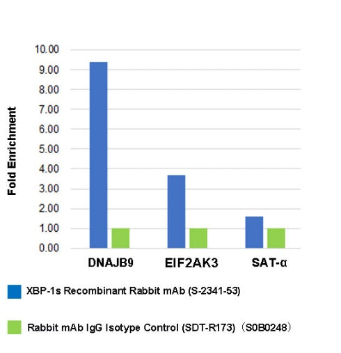
Chromatin immunoprecipitation (ChIP) was performed on HeLa + Tunicamycin (2 ug/ml, 8 h) (+) cross-linked with 1% formaldehyde for 10 min, then chromatin was fragmented by sonication. Parallel reactions used XBP-1s Recombinant Rabbit mAb (S-2341-53) and Rabbit mAb IgG Isotype Control (SDT-R173) at 1:20 for immunoprecipitation. Post- immunoprecipitation, both samples were washed, eluted, and cross-links reversed. Purified DNA was analyzed by qPCR.
qPCR showed the enrichment of DNAJB9, EIF2AK3 and SAT-α in XBP-1s Recombinant Rabbit
mAb (S-2341-53)-immunoprecipitated sample.