WB result of WWP1 Recombinant Rabbit mAb
Primary antibody: WWP1 Recombinant Rabbit mAb at 1/1000 dilution
Lane 1: JAR whole cell lysate 20 µg
Lane 2: MDA-MB-231 whole cell lysate 20 µg
Lane 3: HeLa whole cell lysate 20 µg
Lane 4: A431 whole cell lysate 20 µg
Lane 5: HepG2 whole cell lysate 20 µg
Lane 6: MCF7 whole cell lysate 20 µg
Lane 7: LNCaP whole cell lysate 20 µg
Low exprseeion control: JAR whole cell lysate
Secondary antibody: Goat Anti- rabbit IgG, (H+L), HRP conjugated at 1/10000 dilution
Predicted MW: 105 kDa
Observed MW: 125 kDa
This blot was developed with high sensitivity substrate
WWP1 Recombinant Rabbit mAb (S-4412-25)
WWP1 Recombinant Rabbit mAb (S-4412-25)
Price:
Regular price
$100 USD
Regular price
Sale price
$100 USD
Unit price
per
For shipping services or bulk orders, you may request a quotation.
Secure checkout with

View full details
Product Details
Product Details
Product Specification
| Host | Rabbit |
| Antigen | WWP1 |
| Synonyms | NEDD4-like E3 ubiquitin-protein ligase WWP1; Atrophin-1-interacting protein 5 (AIP5); HECT-type E3 ubiquitin transferase WWP1; TGIF-interacting ubiquitin ligase 1 (Tiul1); WW domain-containing protein 1 |
| Immunogen | Synthetic Peptide |
| Location | Cytoplasm, Nucleus, Cell membrane |
| Accession | Q9H0M0 |
| Clone Number | S-4412-25 |
| Antibody Type | Recombinant mAb |
| Isotype | IgG |
| Application | WB, IHC-P |
| Reactivity | Hu, Ms, Rt |
| Purification | Protein A |
| Concentration | 0.5 mg/ml |
| Conjugation | Unconjugated |
| Physical Appearance | Liquid |
| Storage Buffer | PBS, 40% Glycerol, 0.05% BSA, 0.02% sodium azide |
| Stability & Storage | 12 months from date of receipt / reconstitution, -20 °C as supplied |
Dilution
| application | dilution | species |
| WB | 1:500-1:1000 | Hu, Ms, Rt |
| IHC-P | 1:500 | Hu |
Background
WWP1, full name WW domain-containing E3 ubiquitin ligase 1, is a member of the NEDD4-like protein family composed of 922 amino acids with a molecular weight of approximately 110 kDa. Its protein structure, from the N-terminus to the C-terminus, consists of a C2 domain responsible for membrane localization, four tandem WW protein-interaction domains (responsible for recognizing PY motifs on substrate proteins), and a C-terminal HECT domain responsible for catalyzing ubiquitin transfer. Functionally, the E3 ligase activity of WWP1 is precisely regulated by a "multi-lock" mechanism: in its basal state, the WW domains bind to the HECT catalytic domain to form an autoinhibitory conformation, which is relieved upon phosphorylation or binding to activating proteins, thereby catalyzing the ubiquitination and degradation of various substrates. Its key substrates include SMAD proteins that regulate the TGF-β signaling pathway, tumor suppressors such as p63 and KLF5, and connexin 43 which affects cardiac function. Through the degradation of these critical proteins, WWP1 is broadly involved in embryonic development, immune regulation, viral infection, and tumorigenesis, while dysregulation or cancer-associated mutations of WWP1 are closely linked to various neurological disorders including Troyer syndrome, cardiac diseases, and multiple types of human cancers.
Picture
Picture
Western Blot
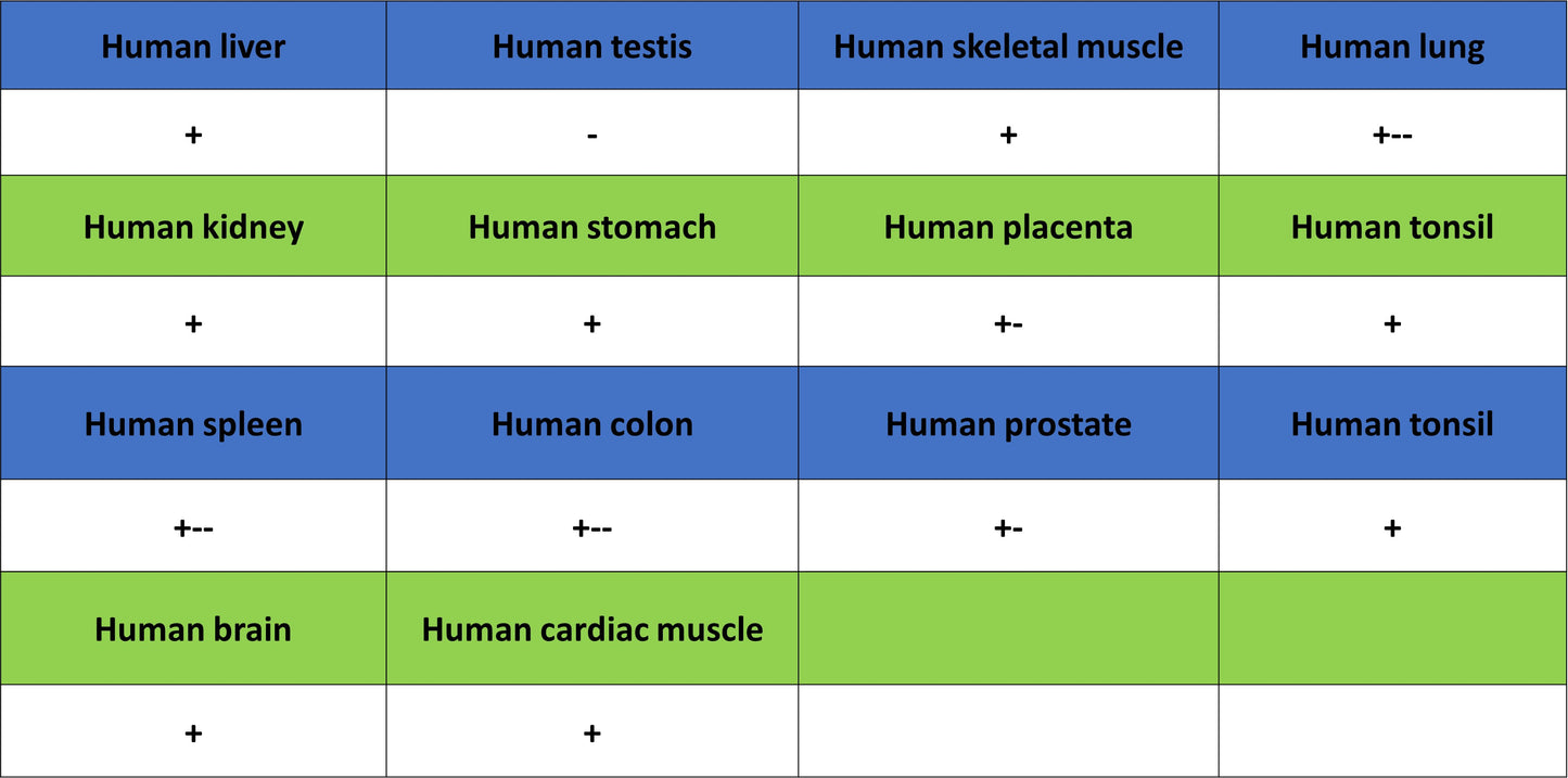
Immunohistochemistry
IHC shows positive staining in paraffin-embedded human cerebral cortex. Anti-WWP1 antibody was used at 1/500 dilution, followed by a HRP Polymer for Rabbit IgG (ready to use). Counterstained with hematoxylin. Heat mediated antigen retrieval with Tris/EDTA buffer pH9.0 was performed before commencing with IHC staining protocol.
IHC shows positive staining in paraffin-embedded human cardiac muscle. Anti-WWP1 antibody was used at 1/500 dilution, followed by a HRP Polymer for Rabbit IgG (ready to use). Counterstained with hematoxylin. Heat mediated antigen retrieval with Tris/EDTA buffer pH9.0 was performed before commencing with IHC staining protocol.
IHC shows positive staining in paraffin-embedded human liver. Anti-WWP1 antibody was used at 1/500 dilution, followed by a HRP Polymer for Rabbit IgG (ready to use). Counterstained with hematoxylin. Heat mediated antigen retrieval with Tris/EDTA buffer pH9.0 was performed before commencing with IHC staining protocol.
IHC shows positive staining in paraffin-embedded human skeletal muscle. Anti-WWP1 antibody was used at 1/500 dilution, followed by a HRP Polymer for Rabbit IgG (ready to use). Counterstained with hematoxylin. Heat mediated antigen retrieval with Tris/EDTA buffer pH9.0 was performed before commencing with IHC staining protocol.
IHC shows positive staining in paraffin-embedded human breast cancer. Anti-WWP1 antibody was used at 1/500 dilution, followed by a HRP Polymer for Rabbit IgG (ready to use). Counterstained with hematoxylin. Heat mediated antigen retrieval with Tris/EDTA buffer pH9.0 was performed before commencing with IHC staining protocol.
IHC shows positive staining in paraffin-embedded human colon cancer. Anti-WWP1 antibody was used at 1/500 dilution, followed by a HRP Polymer for Rabbit IgG (ready to use). Counterstained with hematoxylin. Heat mediated antigen retrieval with Tris/EDTA buffer pH9.0 was performed before commencing with IHC staining protocol.
IHC shows positive staining in paraffin-embedded human pancreatic cancer. Anti-WWP1 antibody was used at 1/500 dilution, followed by a HRP Polymer for Rabbit IgG (ready to use). Counterstained with hematoxylin. Heat mediated antigen retrieval with Tris/EDTA buffer pH9.0 was performed before commencing with IHC staining protocol.