WB result of VGLUT2 Recombinant Rabbit mAb
Primary antibody: VGLUT2 Recombinant Rabbit mAb at 1/1000 dilution
Lane 1: mouse spleen lysate 20 µg
Lane 2: mouse brain lysate 20 µg
Negative control: mouse spleen lysate
Secondary antibody: Goat Anti- rabbit IgG, (H+L), HRP conjugated at 1/10000 dilution
Predicted MW: 65-70 kDa
Observed MW: 65 kDa
VGLUT2 Recombinant Rabbit mAb (S-4399-14)
VGLUT2 Recombinant Rabbit mAb (S-4399-14)
Price:
Regular price
$100 USD
Regular price
Sale price
$100 USD
Unit price
per
For shipping services or bulk orders, you may request a quotation.
Secure checkout with

View full details
Product Details
Product Details
Product Specification
| Host | Rabbit |
| Antigen | VGLUT2 |
| Synonyms | Vesicular glutamate transporter 2; Differentiation-associated BNPI; Differentiation-associated Na(+)-dependent inorganic phosphate cotransporter; Solute carrier family 17 member 6; Dnpi; Vglut2; Slc17a6 |
| Immunogen | Synthetic Peptide |
| Location | Cell membrane, Synapse |
| Accession | Q8BLE7 |
| Clone Number | S-4399-14 |
| Antibody Type | Recombinant mAb |
| Isotype | IgG |
| Application | WB, IHC-P |
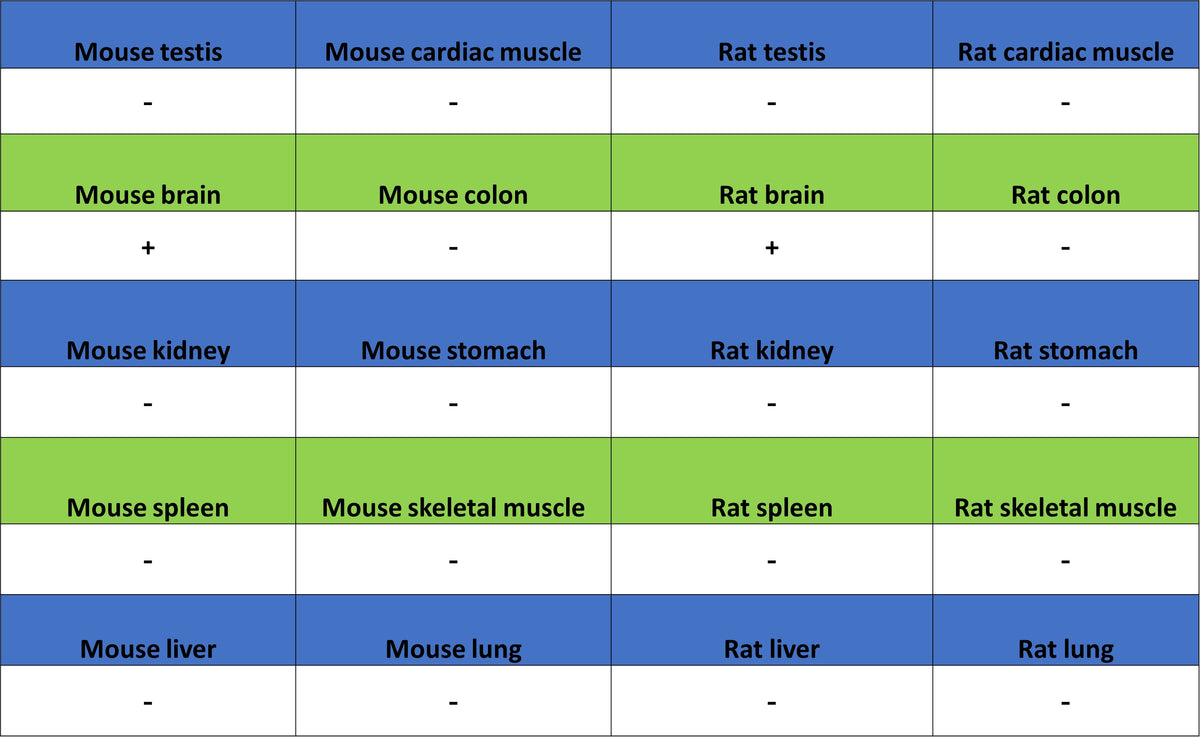
| Reactivity | Ms, Rt |
| Positive Sample | mouse brain, rat brian |
| Purification | Protein A |
| Concentration | 0.5 mg/ml |
| Conjugation | Unconjugated |
| Physical Appearance | Liquid |
| Storage Buffer | PBS, 40% Glycerol, 0.05% BSA, 0.02% sodium azide |
| Stability & Storage | 12 months from date of receipt / reconstitution, -20 °C as supplied |
Dilution
| application | dilution | species |
| WB | 1:1000 | Ms, Rt |
| IHC-P | 1:1000 | Ms, Rt |
Background
VGLUT2 (Vesicular Glutamate Transporter 2), also known as SLC17A6 or DNPI, is a synaptic vesicle protein that mediates the sodium-dependent uptake of glutamate into presynaptic vesicles. As a specific marker for glutamatergic neurons, it plays a crucial role in excitatory neurotransmission and is considered a potential therapeutic target for neurological disorders involving excessive glutamate signaling, such as epilepsy, chronic pain, and neurodegenerative diseases.
Picture
Picture
Western Blot
WB result of VGLUT2 Recombinant Rabbit mAb
Primary antibody: VGLUT2 Recombinant Rabbit mAb at 1/1000 dilution
Lane 1: rat spleen lysate 20 µg
Lane 2: rat brain lysate 20 µg
Negative control: rat spleen lysate
Secondary antibody: Goat Anti- rabbit IgG, (H+L), HRP conjugated at 1/10000 dilution
Predicted MW: 65-70 kDa
Observed MW: 65 kDa
Immunohistochemistry
IHC shows positive staining in paraffin-embedded mouse cerebral cortex. Anti-VGLUT2 antibody was used at 1/1000 dilution, followed by a HRP Polymer for Mouse & Rabbit IgG (ready to use). Counterstained with hematoxylin. Heat mediated antigen retrieval with Tris/EDTA buffer pH9.0 was performed before commencing with IHC staining protocol.
Negative control: IHC shows negative staining in paraffin-embedded mouse spleen. Anti-VGLUT2 antibody was used at 1/1000 dilution, followed by a HRP Polymer for Mouse & Rabbit IgG (ready to use). Counterstained with hematoxylin. Heat mediated antigen retrieval with Tris/EDTA buffer pH9.0 was performed before commencing with IHC staining protocol.
IHC shows positive staining in paraffin-embedded rat cerebral cortex. Anti-VGLUT2 antibody was used at 1/1000 dilution, followed by a HRP Polymer for Mouse & Rabbit IgG (ready to use). Counterstained with hematoxylin. Heat mediated antigen retrieval with Tris/EDTA buffer pH9.0 was performed before commencing with IHC staining protocol.
Negative control: IHC shows negative staining in paraffin-embedded rat spleen. Anti-VGLUT2 antibody was used at 1/1000 dilution, followed by a HRP Polymer for Mouse & Rabbit IgG (ready to use). Counterstained with hematoxylin. Heat mediated antigen retrieval with Tris/EDTA buffer pH9.0 was performed before commencing with IHC staining protocol.