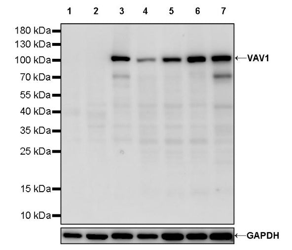
WB result of VAV1 Recombinant Rabbit mAb
Primary antibody: VAV1 Recombinant Rabbit mAb at 1/1000 dilution
Lane 1: A549 whole cell lysate 20 µg
Lane 2: MCF7 whole cell lysate 20 µg
Lane 3: Jurkat whole cell lysate 20 µg
Lane 4: K562 whole cell lysate 20 µg
Lane 5: Raji whole cell lysate 20 µg
Lane 6: Ramos whole cell lysate 20 µg
Lane 7: Molt-4 whole cell lysate 20 µg
Negative control: A549 whole cell lysate
Low expression control: MCF7 whole cell lysate
Secondary antibody: Goat Anti-rabbit IgG, (H+L), HRP conjugated at 1/10000 dilution
Predicted MW: 98 kDa
Observed MW: 105 kDa
This blot was developed with high sensitivity substrate
VAV1 Recombinant Rabbit mAb (S-2499-37)
VAV1 Recombinant Rabbit mAb (S-2499-37)
Price:
Regular price
$170 USD
Regular price
Sale price
$170 USD
Unit price
per
For shipping services or bulk orders, you may request a quotation.
Secure checkout with

View full details
Product Details
Product Details
Product Specification
| Host | Rabbit |
| Synonyms | Proto-oncogene vav; VAV |
| Immunogen | Recombinant Protein |
| Location | Cytoplasm |
| Accession | P15498 |
| Clone Number | S-2499-37 |
| Antibody Type | Recombinant mAb |
| Isotype | IgG |
| Application | WB |
| Reactivity | Hu |
| Purification | Protein A |
| Concentration | 0.5 mg/ml |
| Conjugation | Unconjugated |
| Physical Appearance | Liquid |
| Storage Buffer | PBS, 40% Glycerol, 0.05% BSA, 0.03% Proclin 300 |
| Stability & Storage | 12 months from date of receipt / reconstitution, -20 °C as supplied |
Dilution
| application | dilution | species |
| WB | 1:1000 | Hu |
Background
VAV1 is a 95-kDa multidomain guanine-nucleotide exchange factor (GEF) that is expressed predominantly in hematopoietic cells; it consists of an N-terminal calponin-homology (CH) domain, an acidic region that is phosphorylated by tyrosine kinases, a Dbl-homology (DH) domain responsible for Rho/Rac GEF activity, a Pleckstrin-homology (PH) domain for membrane targeting, two SH3 domains flanking a single SH2 domain, and a C-terminal SH3 domain that mediates protein–protein interactions. Upon engagement of T- and B-cell antigen receptors, VAV1 is rapidly tyrosine-phosphorylated by Src and Syk family kinases, leading to its recruitment to the plasma membrane where it catalyzes GDP-to-GTP exchange on Rac1, Rac2, and RhoG, thereby activating downstream pathways controlling actin cytoskeletal reorganization, integrin clustering, immune-synapse formation, calcium flux, and transcriptional programs via NF-κB, NFAT, and AP-1. In addition to its canonical GEF function, phosphorylated VAV1 serves as a scaffold that binds adaptors such as SLP-76 and LAT to propagate signaling complexes, and it participates in negative feedback through interactions with ubiquitin ligases like c-Cbl. Gain-of-function mutations or overexpression of VAV1 are implicated in T-cell acute lymphoblastic leukemia, peripheral T-cell lymphomas, and neuroblastoma, whereas loss-of-function variants cause combined immunodeficiency with impaired T-cell development and function, highlighting its central role in both adaptive immunity and oncogenesis.
Picture
Picture
Western Blot