WB result of Upf1 Rabbit Polyclonal Antibody
Primary antibody: Upf1 Recombinant Rabbit mAb at 1/1000 dilution
Lane 1: SH-SY5Y whole cell lysate 20 µg
Lane 2: Raji whole cell lysate 20 µg
Lane 3: SK-OV-3 whole cell lysate 20 µg
Lane 4: HeLa whole cell lysate 20 µg
Lane 5: HEK-293 whole cell lysate 20 µg
Lane 6: HuT 78 whole cell lysate 20 µg
Lane 7: Hep G2 whole cell lysate 20 µg
Lane 8: RD whole cell lysate 20 µg
Secondary antibody: Goat Anti-Rabbit IgG (H+L), HRP conjugated at 1/10000 dilution
Predicted MW: 124 kDa
Observed MW: 124 kDa
This blot was developed with high sensitivity substrate
Upf1 Rabbit Polyclonal Antibody
Upf1 Rabbit Polyclonal Antibody
Price:
Regular price
$100 USD
Regular price
Sale price
$100 USD
Unit price
per
For shipping services or bulk orders, you may request a quotation.
Secure checkout with

View full details
Product Details
Product Details
Product Specification
| Host | Rabbit |
| Antigen | UPF1 |
| Synonyms | Regulator of nonsense transcripts 1; ATP-dependent helicase RENT1; Double-stranded nucleic acid 5'-3' helicase UPF1; Nonsense mRNA reducing factor 1Imported (NORF1Imported); Up-frameshift suppressor 1 homolog (hUpf1); KIAA0221; RENT1 |
| Immunogen | Synthetic Peptide |
| Location | Cytoplasm, Nucleus |
| Accession | Q92900 |
| Antibody Type | Polyclonal antibody |
| Isotype | IgG |
| Application | WB, IHC-P, ICC |
| Reactivity | Hu, Ms, Rt |
| Positive Sample | SH-SY5Y, Raji, SK-OV-3, HeLa, HEK-293, HuT 28, HepG2, RD, NIH/3T3, C2C12, mouse liver, C6, rat liver |
| Predicted Reactivity | Zf |
| Purification | Immunogen Affinity |
| Concentration | 0.5 mg/ml |
| Conjugation | Unconjugated |
| Physical Appearance | Liquid |
| Storage Buffer | PBS, 40% Glycerol, 0.05% BSA, 0.02% sodium azide |
| Stability & Storage | 12 months from date of receipt / reconstitution, -20 °C as supplied |
Dilution
| application | dilution | species |
| WB | 1:1000 | Hu, Ms, Rt |
| IHC-P | 1:500 | Hu, Ms, Rt |
| ICC | 1:100 | Hu, Ms, Rt |
Background
UPF1 (Up-Frameshift Mutant 1) is the core regulatory factor of the nonsense-mediated mRNA decay (NMD) pathway, playing a vital "molecular sentinel" role in RNA quality control and exhibiting remarkable functional diversity. Its structure features distinctive modular architecture: the N-terminus contains a zinc finger domain responsible for RNA binding; the central region harbors a unique helicase core belonging to the SF1 superfamily of DNA/RNA helicases, whose helicase activity not only depends on ATP hydrolysis but is also tightly regulated by its own phosphorylation status; and the C-terminus participates in protein-protein interactions. In the classical NMD pathway, UPF1 is recruited to the termination codon during the translation termination phase by forming a "SURF" complex with release factors eRF1/eRF3 and SMG1 kinase, where it undergoes critical phosphorylation. Subsequently, its helicase activity cooperates with UPF2 and UPF3B to remodel the mRNP complex, ultimately triggering degradation mechanisms such as decapping and endonucleolytic cleavage, ensuring the clearance of aberrant transcripts containing premature termination codons. However, UPF1's functions extend far beyond this; it is also a key player in DNA replication and telomere maintenance, capable of binding to nascent DNA, promoting replication fork progression, and suppressing homologous recombination, thereby safeguarding genomic stability. In terms of disease relevance, the biological importance of UPF1 is particularly prominent: in tumors, it regulates the expression of various tumor suppressor genes through the NMD pathway, potentially exerting either tumor-suppressive or tumor-promoting dual effects; and UPF1 mutations are the direct cause of hereditary dyskeratosis, a disease characterized by progressive bone marrow failure and pulmonary fibrosis. Additionally, UPF1 also plays important roles in viral infections (such as HIV-1) and inflammatory responses, making it a molecular target with broad intervention potential across multiple pathophysiological processes.
Picture
Picture
Western Blot
WB result of Upf1 Rabbit Polyclonal Antibody
Primary antibody: Upf1 Recombinant Rabbit mAb at 1/1000 dilution
Lane 1: NIH/3T3 whole cell lysate 20 µg
Lane 2: C2C12 whole cell lysate 20 µg
Lane 3: mouse liver lysate 20 µg
Lane 4: mouse kidney lysate 20 µg
Negative control: mouse kidney lysate
Secondary antibody: Goat Anti-Rabbit IgG (H+L), HRP conjugated at 1/10000 dilution
Predicted MW: 124 kDa
Observed MW: 124 kDa
This blot was developed with high sensitivity substrate
WB result of Upf1 Rabbit Polyclonal Antibody
Primary antibody: Upf1 Recombinant Rabbit mAb at 1/1000 dilution
Lane 1: C6 whole cell lysate 20 µg
Lane 2: rat liver lysate 20 µg
Lane 3: rat kidney lysate 20 µg
Negative control: rat kidney lysate
Secondary antibody: Goat Anti-Rabbit IgG (H+L), HRP conjugated at 1/10000 dilution
Predicted MW: 124 kDa
Observed MW: 124 kDa
This blot was developed with high sensitivity substrate
Immunohistochemistry
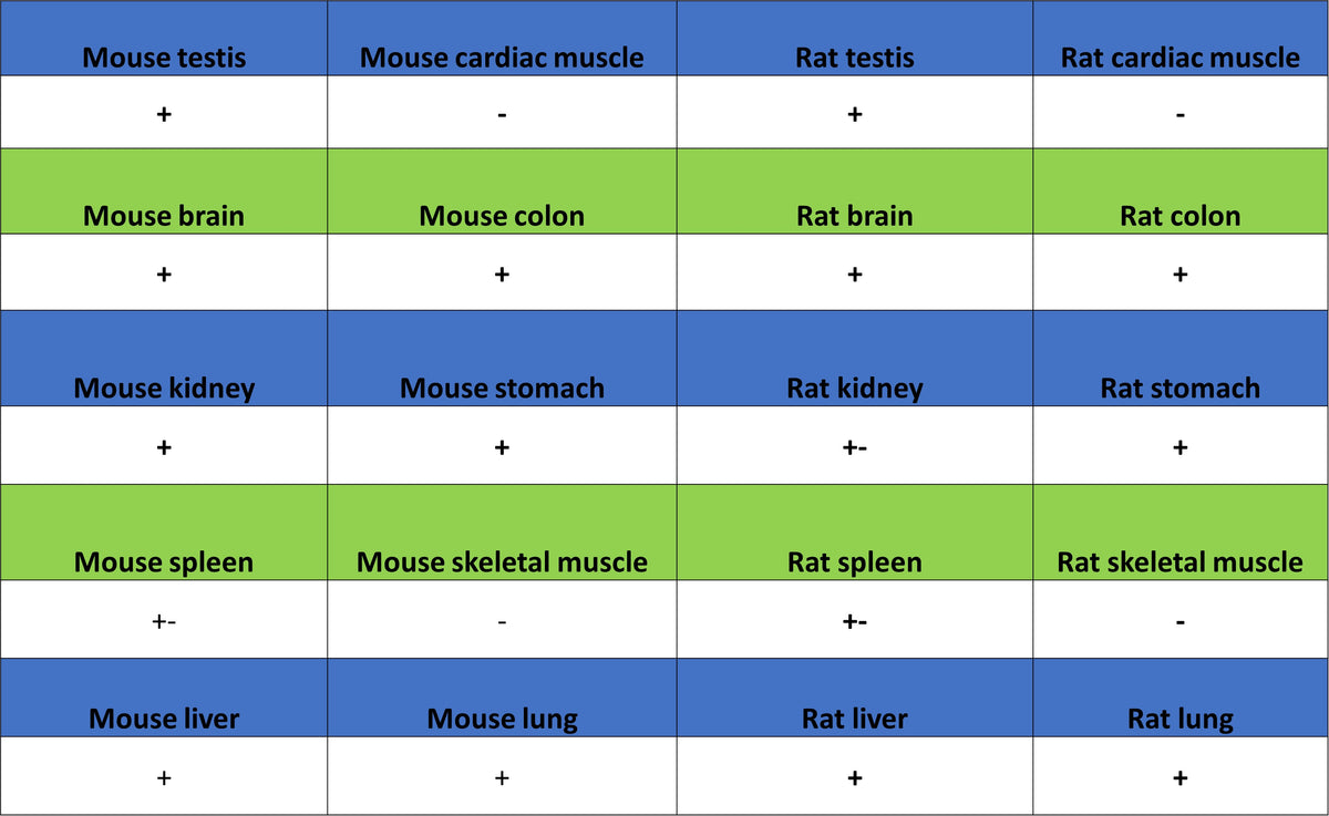
IHC shows positive staining in paraffin-embedded human testis. Anti-Upf1 antibody was used at 1/500 dilution, followed by a HRP Polymer for Rabbit IgG (ready to use). Counterstained with hematoxylin. Heat mediated antigen retrieval with Tris/EDTA buffer pH9.0 was performed before commencing with IHC staining protocol.
IHC shows positive staining in paraffin-embedded human hepatocellular carcinoma. Anti-Upf1 antibody was used at 1/500 dilution, followed by a HRP Polymer for Rabbit IgG (ready to use). Counterstained with hematoxylin. Heat mediated antigen retrieval with Tris/EDTA buffer pH9.0 was performed before commencing with IHC staining protocol.
IHC shows positive staining in paraffin-embedded human pancreatic cancer. Anti-Upf1 antibody was used at 1/500 dilution, followed by a HRP Polymer for Rabbit IgG (ready to use). Counterstained with hematoxylin. Heat mediated antigen retrieval with Tris/EDTA buffer pH9.0 was performed before commencing with IHC staining protocol.
IHC shows positive staining in paraffin-embedded mouse cerebral cortex. Anti-Upf1 antibody was used at 1/500 dilution, followed by a HRP Polymer for Rabbit IgG (ready to use). Counterstained with hematoxylin. Heat mediated antigen retrieval with Tris/EDTA buffer pH9.0 was performed before commencing with IHC staining protocol.
IHC shows positive staining in paraffin-embedded rat testis. Anti-Upf1 antibody was used at 1/500 dilution, followed by a HRP Polymer for Rabbit IgG (ready to use). Counterstained with hematoxylin. Heat mediated antigen retrieval with Tris/EDTA buffer pH9.0 was performed before commencing with IHC staining protocol.
Immunocytochemistry
ICC shows positive staining in Raji cells. Anti-Upf1 antibody was used at 1/100 dilution (Green) and incubated overnight at 4°C. Goat polyclonal Antibody to Rabbit IgG - H&L (Alexa Fluor® 488) was used as secondary antibody at 1/1000 dilution. The cells were fixed with 4% PFA and permeabilized with 0.1% PBS-Triton X-100. Nuclei were counterstained with DAPI (Blue). Counterstain with tubulin (Red).
ICC shows positive staining in NIH/3T3 cells. Anti-Upf1 antibody was used at 1/100 dilution (Green) and incubated overnight at 4°C. Goat polyclonal Antibody to Rabbit IgG - H&L (Alexa Fluor® 488) was used as secondary antibody at 1/1000 dilution. The cells were fixed with 4% PFA and permeabilized with 0.1% PBS-Triton X-100. Nuclei were counterstained with DAPI (Blue). Counterstain with tubulin (Red).
ICC shows positive staining in C6 cells. Anti-Upf1 antibody was used at 1/100 dilution (Green) and incubated overnight at 4°C. Goat polyclonal Antibody to Rabbit IgG - H&L (Alexa Fluor® 488) was used as secondary antibody at 1/1000 dilution. The cells were fixed with 4% PFA and permeabilized with 0.1% PBS-Triton X-100. Nuclei were counterstained with DAPI (Blue). Counterstain with tubulin (Red).