UniOne® TR-FRET KinActive-TK general Kit
UniOne® TR-FRET KinActive-TK general Kit

Product Details
Product Details
Product Specification
| Host | Human |
| Stability & Storage | -80℃ |
Background
UniOne® TR-FRET THUNDER™ KinActive-TK general Kitis a universal method that uses time-resolved fluorescence resonance energy transfer (TR-FRET) technology to measure tyrosine kinase activity. It is suitable for detecting the activity of various tyrosine kinases and screening inhibitors, and the procedure is simple.
As shown in the figure below, the assay consists of two steps: an enzymatic reaction and detection. During the enzymatic reaction, the reaction mixture includes the kinase, a biotinylated substrate, and ATP. After adding ATP, the kinase phosphorylates the biotinylated TK substrate. In the detection step, the detection reagents are diluted with the detection buffer; the EDTA in the buffer terminates the enzymatic reaction. The Eu-labeled antibody and streptavidin-BAc simultaneously capture the phosphorylated substrate. The TR-FRET signal generated by the two detection reagents bound to the substrate is proportional to the level of substrate phosphorylation, which in turn is proportional to the kinase's phosphorylation activity in the system.

Components
[Product Components and Storage Conditions]
Reagent Name |
Concentration |
100 Tests |
500 Tests |
2,500 Tests |
10,000 Tests |
TK Substrate |
100 μM |
10μL |
50 μL |
250 μL |
1 mL |
SA-BAc |
25× |
20 μL |
100 μL |
500μL |
2 mL |
TK-Antibody-Eu |
25× |
20 μL |
100 μL |
500μL |
2 mL |
supplement component |
2,500nM |
10 μL |
50 μL |
500μL |
2 mL |
5×Enzymatic Buffer |
5× |
0.4 mL |
2 mL |
10mL |
40mL |
1×Detection Buffer |
1× |
2 mL |
10 mL |
50mL |
200 mL |
Note: The product mainly includes biotinylated substrates, detection reagents, and buffers. The buffers can be stored at 4°C, while other components should be stored at −80°C.
Add ingredients (not provided)
Kit components not includedEGFRKinase, enzyme reaction buffer with ddH₂O2Dilute to 1×, then add DTT, MgCl₂2, MnCl₂2Prepare the kinase buffer by adding 1 mM DTT, 5 mM MgCl₂.2, 5 mM MnCl₂2The kit does not include ATP; the optimal concentration of ATP must be determined in advance through serial dilutions.
Protocol
1. Reagent Preparation
1.1 Before use, thaw all reagents at room temperature (allow them to equilibrate at room temperature for at least 30 minutes). The reaction system in a 384-well plate is 20μL (the reagent volumes for the reaction system are shown in the table). Calculate the required volume for the experiment before preparation and prepare accordingly; the following preparation is for reference only, taking 500 reactions as an example.
During the enzymatic reaction step, various reagents are diluted with the prepared 1×Enzymatic BufferFor detection reagents, use the provided1×Detection Bufferfor configuration. It is not recommended to store working solutions of various components after preparation; they must be used immediately. To prevent degradation, kinase working solutions should be kept on ice during the experiment.
Components |
Preparation Method |
1×Enzymatic Buffer |
Dilute with ddH2O to5×Enzymatic BufferDilute to 1×, addsupplement component(The concentration needs to be pre-diluted in gradients to determine the optimal usage concentration), DTT, MgCl2, MnCl2Prepare into 1×Enzymatic Buffer. |
1×Detection Buffer |
1×Detection Buffer |
Inhibitor |
Use 1×Enzymatic BufferDilute the compound to prepare a working solution at 2.5 times the final concentration required for the enzymatic reaction step (10 μL). |
TK Substrate |
Dilute the TK Substrate stock solution with 1× Enzymatic Buffer to prepare a5×TK Substrateworking solution,whose concentration is five times the final TK Substrate concentration needed for the enzymatic reaction step.ATPDilute the ATP stock solution with 1× Enzymatic Buffer to prepare a |
ATP |
Dilute the ATP stock solution with 1× Enzymatic Buffer to prepare a5× ATP working solution; the concentration of this working solution is five times the final ATP concentration required for the enzymatic reaction step. (The ATP usage concentration needs to be pre-diluted in gradients to determine the optimal concentration.) |
SA-BAc |
Using1×Detection BufferDilute according to the specified ratio to obtain a 1× working solution. |
BTK-Antibody-Eu |
Using1Detection BufferDilute according to the dilution factor to obtain a 1× working solution. |
1.2Sample to be tested(Inhibitor) gradient dilution
Dilute the compound with 1× Enzymatic Buffer, and determine the dilution factor and gradient based on the consistent inhibitory effect of the inhibitor.
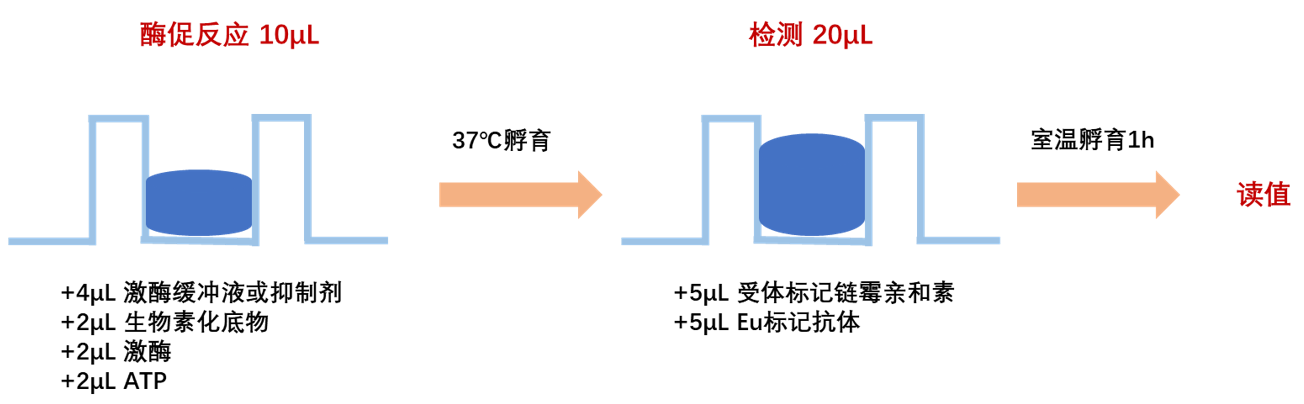
2. Reaction system
As shown in the figure, taking a 384-well plate as an example, add the inhibitor, substrate, kinase, and ATP, then incubate at room temperature or 37°C (the incubation time and temperature for the enzymatic reaction should be optimized according to the target kinase) for 30 minutes (in the enzymatic reaction system, it is recommended that the DMSO concentration does not exceed 2%). After the reaction, add the detection reagent; the EDTA in the detection buffer will terminate the enzymatic reaction. Incubate at room temperature for 1 hour, then read the values on the instrument.
The experiment should include the following control wells:
(1) Negative control: non-specific signal, used to calculate the specific signal value of the reaction system.
(2) Buffer control: to eliminate the influence of the buffer on the signal value.
Enzymatic reaction |
Control |
||||
Components |
Experimental well |
Negative control |
Eu-labeled antibody |
Buffer |
|
Inhibitor/kinase buffer |
4 μL |
4 μL1×Enzymatic Buffer |
10 μL |
10 μL |
1.Enzymatic reaction (10 μL) incubate at room temperature or 37℃ for 30 min. |
TK |
2 μL |
2 μL |
|||
TK Substrate |
2 μL |
2 μL1×Enzymatic Buffer |
|||
ATP |
2 μL |
2 μL |
|||
SA-BAc |
5 μL |
5 μL |
5 μL 1×Detection Buffer |
10 μL |
2.Detection steps Incubate at room temperature for 1h. |
Eu-labeled antibody |
5 μL |
5 μL |
5 μL |
||
(3)Eu-labeled antibody control: used to verify the normal signal value of the Eu-labeled antibody at 620 nM.
[Detection]
Detect on a compatible TR-FRET microplate reader (excitation at 320 nm, emission wavelengths at 620 nm and 665 nm).
[Result Calculation]
1) Ratio:
Ratio = (665/620)×10000
2) Net signal:
Net signal= (Std-NC)/NC×100
Std: the ratio of sample and control
NC: the ratio ofnegative control
3)CV (%):
CV (%) = Standard Deviation / Mean Ratio × 100%
Note: Recommendedmicroplate (384-well plate, white, shallow wells), item number: abs7311;
[Kinase Experiment Optimization]
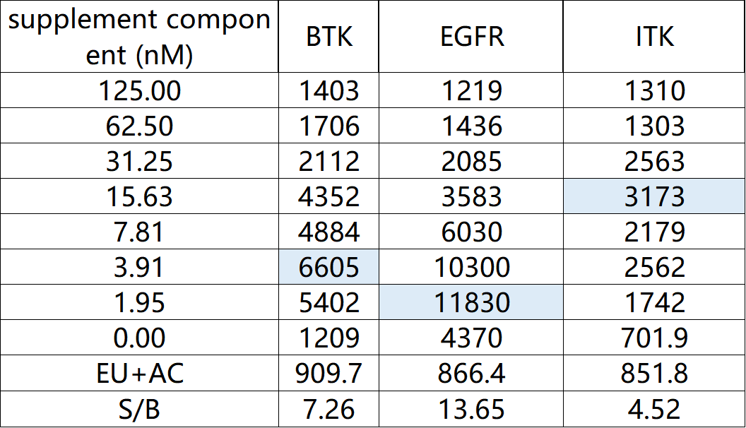
1)supplement componentconcentration optimization
Some tyrosine kinases may require the addition ofsupplement componentreagents to kinase buffer 1X in order to achieve optimal enzyme activity. This step can determine the bestsupplement componentConcentration—refers to the concentration corresponding to when the signal reaches its maximum value of 80%. Prepare a series of solutions with different concentrations.supplement componentof kinase buffer at 1X (recommended range from 125 nM to 0 nM).
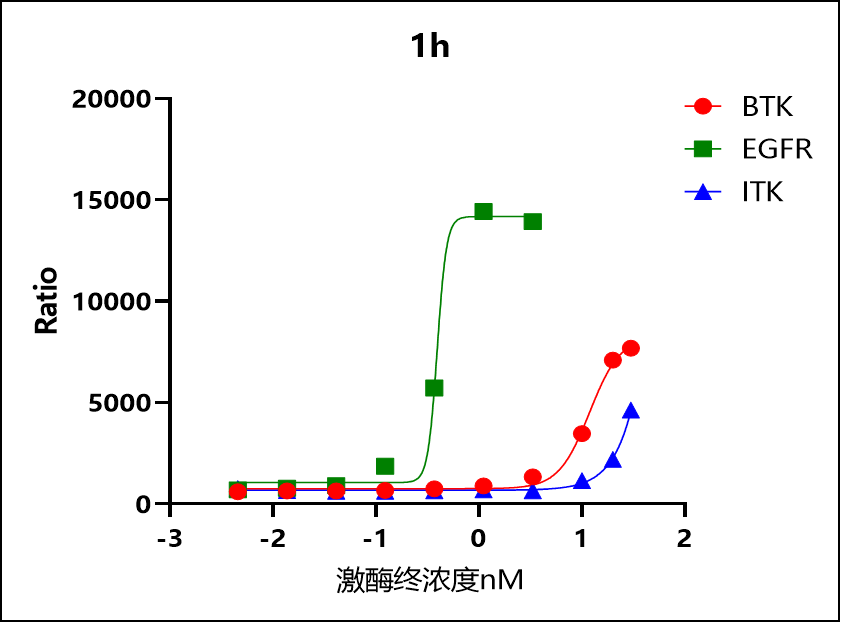
2)Kinase concentration optimization
This step is primarily aimed at determining the optimal enzyme concentration (at which the signal reaches 80% of its maximum). A balance should be struck between high detection signals and enzyme consumption. Use a fixed concentration of TK-substrate-biotin (e.g., 1 µM) and ATP (e.g., 100 µM), perform gradient dilutions of the enzyme, and test them (recommended enzyme concentrations are 10, 2, 1, and 0 ng/well; these can also be adjusted flexibly based on preliminary data). Allow the enzymatic reaction to proceed for 30 minutes. The recommended biotin/streptavidin ratio is 8:1.
3)Kinetic studies
The kinetic properties of the enzyme depend on the concentrations of the kinase and the substrate. In incubation time studies, a fixed concentration of kinase (determined from previous experiments), ATP (100 μM), and substrate (1 µM) should be used; reaction termination at different time points is achieved by adding the detection reagent at 1, 2, 5, 10, 30, and 60 minutes. The recommended ratio of biotin to streptavidin is 8:1.
4)Substrate concentration optimization
Use the optimal enzyme concentration and a saturated ATP concentration (100 µM). It is recommended to test different concentrations of the TK substrate-biotin, with a concentration gradient ranging from 2 µM to 1 nM (two-fold serial dilutions). The kinase reaction should be terminated at the previously determined optimal incubation time. During the detection step, adjust the streptavidin concentration according to the concentration of the TK substrate-biotin to ensure that the biotin/streptavidin ratio remains at 8:1. Since background signals may increase as the streptavidin concentration rises, for eachstreptavidina negative control must be set up.
5)ATP concentration optimization
This step optimizes the ATP concentration, requiring the use of the optimal enzyme concentration and a saturated TK substrate concentration (1 µM). It is recommended to test ATP concentrations ranging from 300 µM to 1.7 nM (using threefold serial dilutions). At the optimal incubation time, the kinase reaction is terminated by adding the detection reagent. The recommended ratio of biotin to streptavidin is 8:1.
6)Biotin/Streptavidin Ratio Optimization
Optimizing the biotin/streptavidin ratio is a critical step that can significantly enhance signal intensity. Prepare streptavidin solutions with different biotin/streptavidin ratios (2/1, 4/1, 8/1); in the experiment, use the optimal enzyme concentration, ATP concentration, and substrate concentration for detection; meanwhile, each streptavidin concentration should be accompanied by its corresponding negative control, as this reagent directly affects the background value.
7)IC50 determination
Kinase activity must be tested across a broad range of inhibitor concentrations to generate a dose-response curve; this assay is typically performed under previously established optimal assay conditions.