UniOne® TR-FRET Human FcRn Binding Kit
UniOne® TR-FRET Human FcRn Binding Kit

Product Details
Product Details
Product Specification
| Host | Human |
| Stability & Storage | -80℃ |
Background
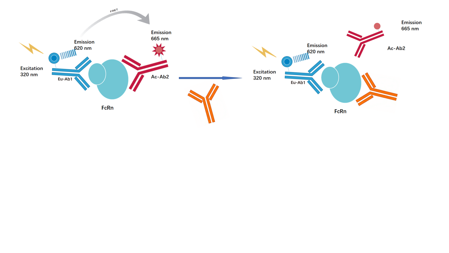
The kit is based on homogeneous time-resolved fluorescence technology (TR-FRET) technology,which is used to assess the interaction between the FcRn and IgG. This method provides a high-throughput approach to identify the ability of antibody drugs to bind to FcRn simply and rapidly.
As shown in the figure below, the FcRn detected by an Eu-labeled anti-Tag1 antibody (TR-FRET donor) interacted with Ac-labeled antibody (TR-FRET receptor), and the excitation of the donor antibody triggers fluorescence resonance energy transfer (FRET) to the receptor antibody, resulting in specific emission of a signal at a wavelength of 665 nm from the receptor antibody. While the antibody drugs can compete with the Ac-labeled antibody, binding with the FcRn protein, resulting in a decrease in the signal at a wavelength of 665 nm. This decrease of specific signal is proportional to the degree of interaction between FcRn and antibody drugs. This homogeneous assay is simple to perform and requires no washing steps.
Components
Composition |
concentration |
100T |
500T |
2500T |
10000T |
Storage |
Tag1-FcRn protein |
25× |
20μL |
100μL |
500μL |
2000μL |
-80℃ |
IgG calibrator |
11.8mg/mL |
5μL |
10μL |
50μL |
200μL |
-80℃ |
Anti-Tag1 Eu antibody |
50× |
5μL |
25μL |
125μL |
500μL |
-80℃ |
IgG Ac antibody |
100× |
5μL |
13μL |
63μL |
250μL |
-80℃ |
Detection buffer |
1× |
4mL |
20mL |
100mL |
400mL |
-80℃ |
Note: Aliquot into appropriate volumes, store at the recommended temperature and avoid repeated freeze-thaw cycle.
Protocol
1、Reagent Preparation
1.1 Compositions equilibration
Before use, equilibrate all reagents at room temperature (allow at least 30 minutes for temperature equilibration). For the 384-well plate assay, use 20 μL of reaction mixture (the reagent quantities are listed in the table below). Calculate the required volume prior to preparation accordingly. The following preparation steps are provided for reference only, using a 500T as an example.
Table 1. Reagent Preparation
Composition |
Preparation |
μL/well |
Tag1-FcRn protein |
Take 100 μL of the 25× Tag1-FcRn protein stock solution, add 2.4mL of 1× Detection buffer to dilute it to 1× concentration, and mix thoroughly for later use. |
5 |
Antibody Mix |
Take 25 μL of the 50× Anti-Tag1 Eu antibody stock solution, add 1225μL of 1× Detection buffer to dilute to 1250 μL, and mix thoroughly; take 13 μL of the 100× IgG Ac antibody stock solution, add 1287 μL of 1× Detection buffer to dilute to 1300 μL, and mix thoroughly; combine the two solutions in a 1:1 ratio to obtain the Antibody Mix. |
5 |
1.2 Gradient dilution of the sample
IgG calibrator as an example, the dilution solution is 1× Detection buffer. To minimize interference from matrix effects, it is recommended to use a solution with the same matrix composition as the sample being tested; additionally, the sample concentration should be adjusted according to its actual situation.
Table 2. IgG calibrator gradient dilution(Adjust according to the actual situation)
IgG calibrator final concentration(μg/ml) |
IgG calibrator preparation concentration(μg/ml) |
Dilution |
|
① |
500 |
1000 |
2μL 11.8mg/mL IgG calibrator +21.6μL 1× Detection buffer |
② |
125 |
250 |
7μL ① +21μL 1× Detection buffer |
③ |
31.25 |
62.5 |
7μL ② +21μL 1× Detection buffer |
④ |
7.813 |
15.625 |
7μL ③ +21μL 1× Detection buffer |
⑤ |
1.953 |
3.906 |
7μL ④ +21μL 1× Detection buffer |
⑥ |
0.488 |
0.977 |
7μL ⑤ +21μL 1× Detection buffer |
⑦ |
0.122 |
0.244 |
7μL ⑥ +21μL 1× Detection buffer |
⑧ |
0.031 |
0.061 |
7μL ⑦+21μL 1× Detection buffer |
Blank |
0 |
0 |
21μL 1× Detection buffer |
2、Sample and control
2.1 Sample:Add 5 μL of Tag1-FcRn protein working solution, 10 μL of the gradient-diluted sample , 500rpm shaken at 25℃ for 30 min,and 5 μL of the Antibody Mix sequentially into the 384-well plate.
2.2 IgG calibrator: Add 5 μL of Tag1-FcRn protein working solution, 10 μL of the gradient-diluted IgG calibrator , 500rpm shaken at 25℃ for 30 min,and 5 μL of the Antibody Mix sequentially into the 384-well plate.
2.3 Blank:Add 5 μL of Tag1-FcRn protein working solution, 10 μL of 1× Detection buffer, 500rpm shaken at 25℃ for 30 min,and 5 μL of the Antibody Mix sequentially into the 384-well plate.
2.4 Negative control: 15μL Detection buffer, and 5 μL of the mixed Antibody Mix sequentially into the 384-well plate.
After adding all samples, centrifuge, seal with a cover membrane, and incubate at room temperature for 1.5 hours.
Table 3. Sample and control spotting process
IgG calibrator |
Sample |
Blank |
Negative control (NC) |
|
10μL gradient-diluted IgG calibrator |
10μL gradient-diluted sample |
10μL 1× Detection buffer |
15μL 1× Detection buffer and 5μL Antibody Mix |
5μL FcRn protein | |||
500rpm shaken at 25℃ for 30 min | |||
5μL Antibody Mix | |||
Seal the plate wells with a sealing film and incubate at 25℃ for 1.5 hours. | |||
3、Read Data
The detection was performed on a TR-FRET microplate reader. The excitation wavelength was 320/340 nm, and the emission wavelengths at 620 nm and 665 nm were detected.
【DATA REDUCTION 】
1) Ratio:
Ratio = (665/620) ×10000
2) Net signal:
Net signal = (Std-NC)/NC×100
Std:the ratio of sample and control
NC: the ratio of negative control
3) CV(%):
CV(%)= Standard Deviation/Mean Ratio × 100%
【Data Example】
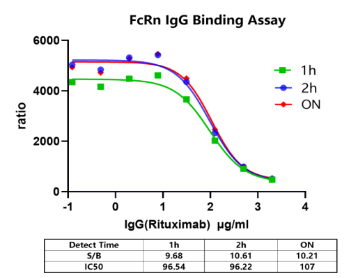
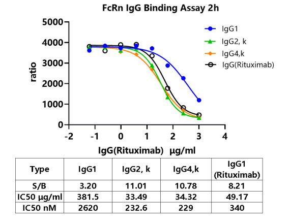
The following data cannot replace experimental results obtained in experiments; they are provided solely for illustrative purposes, and outcomes may vary depending on the plate reader used.
Note: Recommended microplate (384-well plate, white, shallow wells).