WB result of TUG Recombinant Rabbit mAb
Primary antibody: TUG Recombinant Rabbit mAb at 1/1000 dilution
Lane 1: HEK-293 whole cell lysate 20 µg
Lane 2: K562 whole cell lysate 20 µg
Lane 3: HeLa whole cell lysate 20 µg
Lane 4: A549 whole cell lysate 20 µg
Lane 5: 293T whole cell lysate 20 µg
Secondary antibody: Goat Anti- rabbit IgG, (H+L), HRP conjugated at 1/10000 dilution
Predicted MW: 60 kDa
Observed MW: 70 kDa
Tug Recombinant Rabbit mAb (S-4377-21)
Tug Recombinant Rabbit mAb (S-4377-21)
Price:
Regular price
$100 USD
Regular price
Sale price
$100 USD
Unit price
per
For shipping services or bulk orders, you may request a quotation.
Secure checkout with

View full details
Product Details
Product Details
Product Specification
| Host | Rabbit |
| Antigen | Tug |
| Synonyms | Tether containing UBX domain for GLUT4; Alveolar soft part sarcoma chromosomal region candidate gene 1 protein; Alveolar soft part sarcoma locus; Renal papillary cell carcinoma protein 17; UBX domain-containing protein 9; ASPL; RCC17; TUG; UBXD9; UBXN9; ASPSCR1 |
| Immunogen | Synthetic Peptide |
| Location | Cytoplasm, Nucleus, Endoplasmic reticulum |
| Accession | Q9BZE9 |
| Clone Number | S-4377-21 |
| Antibody Type | Recombinant mAb |
| Isotype | IgG |
| Application | WB, IHC-P |
| Reactivity | Hu, Ms, Rt |
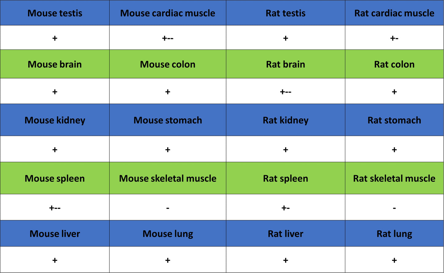
| Positive Sample | HEK-293, K562, HeLa, A549, 293T, NIH/3T3, mouse testis, rat testis, rat heart |
| Purification | Protein A |
| Concentration | 0.5 mg/ml |
| Conjugation | Unconjugated |
| Physical Appearance | Liquid |
| Storage Buffer | PBS, 40% Glycerol, 0.05% BSA, 0.02% sodium azide |
| Stability & Storage | 12 months from date of receipt / reconstitution, -20 °C as supplied |
Dilution
| application | dilution | species |
| WB | 1:1000-1:2000 | Hu, Ms, Rt |
| IHC-P | 1:2000 | Hu, Ms, Rt |
Background
TUG (also known as UBXN9 or Aspscr1) is a multifunctional protein that plays a critical role in intracellular membrane trafficking and glucose metabolism regulation. Its most core function is to serve as the "molecular anchor" for the GLUT4 glucose transporter, tethering GLUT4-containing vesicles to the Golgi matrix in the basal state, thereby preventing their translocation to the plasma membrane. The N-terminus of TUG contains a ubiquitin-like domain (UBL1) that mediates protein-protein interactions, while its C-terminus binds to Golgi matrix proteins, anchoring GLUT4 storage vesicles intracellularly through a "two-end capture" mechanism. Upon insulin stimulation, the Usp25m protease in adipose and muscle cells triggers site-specific cleavage of TUG, generating two functionally independent key fragments: the N-terminal cleavage product TUGUL acts as a novel ubiquitin-like modifier that modifies the KIF5B kinesin motor, driving GLUT4 vesicle transport toward the cell surface; meanwhile, the C-terminal product is released from the Golgi under the action of p97 (VCP) ATPase and translocates to the nucleus, where it binds to transcriptional regulators such as PPARγ and PGC-1α to promote the expression of genes involved in fatty acid oxidation and thermogenesis, thereby coupling glucose uptake with energy metabolism regulation. Beyond this classical insulin-regulated function, recent studies have revealed that TUG protein also localizes to the endoplasmic reticulum-Golgi intermediate compartment (ERGIC), where it is recruited to the ERGIC membrane via its central intrinsically disordered region and is essential for maintaining the structural integrity of this compartment and regulating anterograde trafficking in the early secretory pathway. The functional versatility of TUG makes it an important target for studying insulin resistance, type 2 diabetes, and energy metabolism disorders.
Picture
Picture
Western Blot
WB result of TUG Recombinant Rabbit mAb
Primary antibody: TUG Recombinant Rabbit mAb at 1/1000 dilution
Lane 1: NIH/3T3 whole cell lysate 20 µg
Lane 2: mouse testis lysate 20 µg
Secondary antibody: Goat Anti- rabbit IgG, (H+L), HRP conjugated at 1/10000 dilution
Predicted MW: 60 kDa
Observed MW: 70 kDa
WB result of TUG Recombinant Rabbit mAb
Primary antibody: TUG Recombinant Rabbit mAb at 1/1000 dilution
Lane 1: rat testis lysate 20 µg
Lane 2: rat heart lysate 20 µg
Lane 3: rat skeletal muscle lysate 20 µg
Secondary antibody: Goat Anti- rabbit IgG, (H+L), HRP conjugated at 1/10000 dilution
Predicted MW: 60 kDa
Observed MW: 70 kDa
Immunohistochemistry
IHC shows positive staining in paraffin-embedded human testis. Anti-Tug antibody was used at 1/2000 dilution, followed by a HRP Polymer for Rabbit IgG (ready to use). Counterstained with hematoxylin. Heat mediated antigen retrieval with Tris/EDTA buffer pH9.0 was performed before commencing with IHC staining protocol.
IHC shows positive staining in paraffin-embedded human breast cancer. Anti-Tug antibody was used at 1/2000 dilution, followed by a HRP Polymer for Rabbit IgG (ready to use). Counterstained with hematoxylin. Heat mediated antigen retrieval with Tris/EDTA buffer pH9.0 was performed before commencing with IHC staining protocol.
IHC shows positive staining in paraffin-embedded human lung cancer. Anti-Tug antibody was used at 1/2000 dilution, followed by a HRP Polymer for Rabbit IgG (ready to use). Counterstained with hematoxylin. Heat mediated antigen retrieval with Tris/EDTA buffer pH9.0 was performed before commencing with IHC staining protocol.
IHC shows positive staining in paraffin-embedded human ovarian cancer. Anti-Tug antibody was used at 1/2000 dilution, followed by a HRP Polymer for Rabbit IgG (ready to use). Counterstained with hematoxylin. Heat mediated antigen retrieval with Tris/EDTA buffer pH9.0 was performed before commencing with IHC staining protocol.
IHC shows positive staining in paraffin-embedded human pancreatic cancer. Anti-Tug antibody was used at 1/2000 dilution, followed by a HRP Polymer for Rabbit IgG (ready to use). Counterstained with hematoxylin. Heat mediated antigen retrieval with Tris/EDTA buffer pH9.0 was performed before commencing with IHC staining protocol.
IHC shows positive staining in paraffin-embedded mouse kidney. Anti-Tug antibody was used at 1/2000 dilution, followed by a HRP Polymer for Rabbit IgG (ready to use). Counterstained with hematoxylin. Heat mediated antigen retrieval with Tris/EDTA buffer pH9.0 was performed before commencing with IHC staining protocol.
IHC shows positive staining in paraffin-embedded rat stomach. Anti-Tug antibody was used at 1/2000 dilution, followed by a HRP Polymer for Rabbit IgG (ready to use). Counterstained with hematoxylin. Heat mediated antigen retrieval with Tris/EDTA buffer pH9.0 was performed before commencing with IHC staining protocol.