WB result of Thrombospondin 1 Recombinant Rabbit mAb
Primary antibody: Thrombospondin 1 Recombinant Rabbit mAb at 1/××× dilution
Lane 1: mouse platelet lysate 5 µg
Secondary antibody: Goat Anti-Rabbit IgG, (H+L), HRP conjugated at 1/10000 dilution
Predicted MW: 129 kDa
Observed MW: 140, 180 kDa
Thrombospondin 1 Recombinant Rabbit mAb (S-4153-22)
Thrombospondin 1 Recombinant Rabbit mAb (S-4153-22)
Price:
Regular price
$100 USD
Regular price
Sale price
$100 USD
Unit price
per
For shipping services or bulk orders, you may request a quotation.
Secure checkout with

View full details
Product Details
Product Details
Product Specification
| Host | Rabbit |
| Antigen | Thrombospondin 1 |
| Synonyms | Thrombospondin-1; TSP; TSP1; THBS1 |
| Immunogen | Synthetic Peptide |
| Location | Secreted, Endoplasmic reticulum |
| Accession | P07996 |
| Clone Number | S-4153-22 |
| Antibody Type | Recombinant mAb |
| Isotype | IgG |
| Application | WB, IHC-P |
| Reactivity | Hu, Ms, Rt |
| Positive Sample | mouse platelet, rat platelet |
| Purification | Protein A |
| Concentration | 0.5 mg/ml |
| Conjugation | Unconjugated |
| Physical Appearance | Liquid |
| Storage Buffer | PBS, 40% Glycerol, 0.05% BSA, 0.02% sodium azide |
| Stability & Storage | 12 months from date of receipt / reconstitution, -20 °C as supplied |
Dilution
| application | dilution | species |
| WB | 1:1000-1:2000 | Hu, Ms, Rt |
| IHC-P | 1:2000 | Hu, Ms, Rt |
Background
Thrombospondin-1 (TSP-1) is a multifunctional secreted glycoprotein composed of trimers and belongs to the matricellular protein family within the extracellular matrix. It contains multiple domains that interact with various cell surface receptors (such as CD36, CD47, integrins) and matrix molecules (such as heparin, fibronectin, collagen). Physiologically, TSP-1 is best known as an endogenous angiogenesis inhibitor—it binds to the CD36 receptor, activating p38 MAPK and caspase signaling pathways to directly induce endothelial cell apoptosis, thereby potently suppressing angiogenesis. Additionally, TSP-1 is a key activator of transforming growth factor-beta (TGF-β), releasing active TGF-β through conformational changes to regulate immune responses, fibrosis, and tissue remodeling. Pathologically, dysregulation of TSP-1 expression is associated with various diseases: in the tumor microenvironment, its high expression can inhibit tumor angiogenesis and immune surveillance, though it may also be associated with tumor progression and metastasis. In cardiovascular disease, TSP-1 inhibits nitric oxide signaling via the CD47 pathway, promoting platelet aggregation and thrombosis. In diabetic nephropathy, TSP-1-mediated TGF-β activation drives renal fibrosis. Therefore, TSP-1 serves as a crucial regulatory hub in the extracellular matrix, playing complex and important roles in vascular biology, immune regulation, and fibrotic diseases.
Picture
Picture
Western Blot
WB result of Thrombospondin 1 Recombinant Rabbit mAb
Primary antibody: Thrombospondin 1 Recombinant Rabbit mAb at 1/××× dilution
Lane 1: rat platelet lysate 5 µg
Secondary antibody: Goat Anti-Rabbit IgG, (H+L), HRP conjugated at 1/10000 dilution
Predicted MW: 129 kDa
Observed MW: 140, 180 kDa
Immunohistochemistry
IHC shows positive staining in paraffin-embedded human bone marrow. Anti- Thrombospondin 1 antibody was used at 1/2000 dilution, followed by a HRP Polymer for Rabbit IgG (ready to use). Counterstained with hematoxylin. Heat mediated antigen retrieval with Tris/EDTA buffer pH9.0 was performed before commencing with IHC staining protocol.
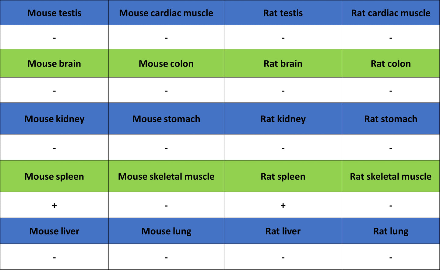
IHC shows positive staining in paraffin-embedded human spleen. Anti- Thrombospondin 1 antibody was used at 1/2000 dilution, followed by a HRP Polymer for Rabbit IgG (ready to use). Counterstained with hematoxylin. Heat mediated antigen retrieval with Tris/EDTA buffer pH9.0 was performed before commencing with IHC staining protocol.
IHC shows positive staining in paraffin-embedded mouse bone marrow. Anti- Thrombospondin 1 antibody was used at 1/2000 dilution, followed by a HRP Polymer for Rabbit IgG (ready to use). Counterstained with hematoxylin. Heat mediated antigen retrieval with Tris/EDTA buffer pH9.0 was performed before commencing with IHC staining protocol.
IHC shows positive staining in paraffin-embedded mouse spleen. Anti- Thrombospondin 1 antibody was used at 1/2000 dilution, followed by a HRP Polymer for Rabbit IgG (ready to use). Counterstained with hematoxylin. Heat mediated antigen retrieval with Tris/EDTA buffer pH9.0 was performed before commencing with IHC staining protocol.
IHC shows positive staining in paraffin-embedded rat bone marrow. Anti- Thrombospondin 1 antibody was used at 1/2000 dilution, followed by a HRP Polymer for Rabbit IgG (ready to use). Counterstained with hematoxylin. Heat mediated antigen retrieval with Tris/EDTA buffer pH9.0 was performed before commencing with IHC staining protocol.
IHC shows positive staining in paraffin-embedded rat spleen. Anti- Thrombospondin 1 antibody was used at 1/2000 dilution, followed by a HRP Polymer for Rabbit IgG (ready to use). Counterstained with hematoxylin. Heat mediated antigen retrieval with Tris/EDTA buffer pH9.0 was performed before commencing with IHC staining protocol.