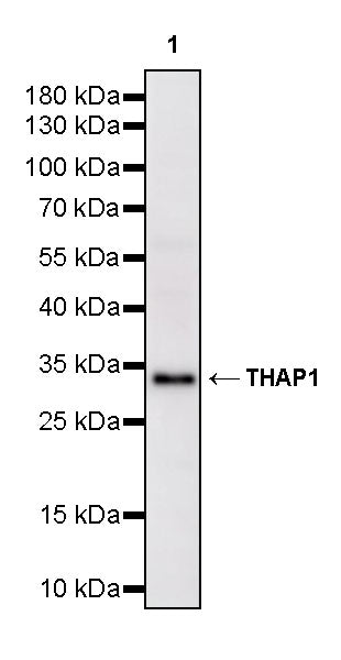
WB result of THAP1 Recombinant Rabbit mAb
Primary antibody: THAP1 Recombinant Rabbit mAb at 1/1000 dilution
Lane 1: 293F transfected with empty vector whole cell lysate 5 µg
Lane 2: 293F transfected with THAP1-Myc-His fusion protein whole cell lysate 5 µg
Secondary antibody: Goat Anti-rabbit IgG (H+L), HRP conjugated at 1/10000 dilution
Predicted MW: 25 kDa
Observed MW: 34 kDa
THAP1 Recombinant Rabbit mAb (S-4023-36)
THAP1 Recombinant Rabbit mAb (S-4023-36)
Price:
Regular price
$100 USD
Regular price
Sale price
$100 USD
Unit price
per
For shipping services or bulk orders, you may request a quotation.
Secure checkout with

View full details
Product Details
Product Details
Product Specification
| Host | Rabbit |
| Antigen | THAP1 |
| Synonyms | THAP domain-containing protein 1 |
| Immunogen | Synthetic Peptide |
| Location | Nucleus |
| Accession | Q9NVV9 |
| Clone Number | S-4023-36 |
| Antibody Type | Recombinant mAb |
| Isotype | IgG |
| Application | WB |
| Reactivity | Hu, Ms, Rt |
| Purification | Protein A |
| Concentration | 2 mg/ml |
| Conjugation | Unconjugated |
| Physical Appearance | Liquid |
| Storage Buffer | PBS, 40% Glycerol, 0.05% BSA, 0.02% sodium azide |
| Stability & Storage | 12 months from date of receipt / reconstitution, -20 °C as supplied |
Dilution
| application | dilution | species |
| WB | 1:1000 | Hu, Ms, Rt |
Background
THAP1 is a transcription factor encoded by the THAP1 gene, containing a characteristic THAP domain (a C2-CH type zinc finger structure) at its N-terminus, which enables specific recognition and binding to DNA sequences to regulate the expression of downstream genes. This protein is the causative gene for DYT6 type (DYT-THAP1), an autosomal dominant inherited form of dystonia, with at least 75 associated mutations having been reported to date, most of which are concentrated in the THAP domain and the coiled-coil domain. Recent studies have revealed a key function of THAP1: it directly binds to and activates the promoter of the PSMB5 gene, which encodes the proteasome core catalytic subunit β5, essential for maintaining basal proteasome activity and intracellular protein homeostasis. Loss of THAP1 function leads to decreased PSMB5 expression, impaired proteasome assembly, and accumulation of ubiquitinated proteins, which may represent an important molecular mechanism underlying the pathogenesis of DYT6 dystonia. In addition, THAP1 possesses a self-negative feedback regulatory mechanism, whereby its own protein product can bind to its own promoter region to inhibit its own transcription.
Picture
Picture
Western Blot
WB result of THAP1 Recombinant Rabbit mAb
Primary antibody: THAP1 Recombinant Rabbit mAb at 1/1000 dilution
Lane 1: HeLa whole cell lysate 20 µg
Lane 2: HEK-293 whole cell lysate 20 µg
Lane 3: Hep G2 whole cell lysate 20 µg
Lane 4: A549 whole cell lysate 20 µg
Secondary antibody: Goat Anti-rabbit IgG (H+L), HRP conjugated at 1/10000 dilution
Predicted MW: 25 kDa
Observed MW: 34 kDa
WB result of THAP1 Recombinant Rabbit mAb
Primary antibody: THAP1 Recombinant Rabbit mAb at 1/1000 dilution
Lane 1: mouse brain lysate 20 µg
Secondary antibody: Goat Anti-rabbit IgG (H+L), HRP conjugated at 1/10000 dilution
Predicted MW: 25 kDa
Observed MW: 34 kDa
WB result of THAP1 Recombinant Rabbit mAb
Primary antibody: THAP1 Recombinant Rabbit mAb at 1/1000 dilution
Lane 1: rat brain lysate 20 µg
Secondary antibody: Goat Anti-rabbit IgG (H+L), HRP conjugated at 1/10000 dilution
Predicted MW: 25 kDa
Observed MW: 34 kDa