Th17 Polarization Kit, Human
Th17 Polarization Kit, Human

Product Details
Product Details
Product Specification
| Species | Human | ||||
| Reconstitution |
|
||||
| Stability & Storage |
|
Components
Component |
Formulation |
Reference dosage |
10ml System(S Size) |
100ml System(M Size) |
1000ml System(L Size) |
IL-6 |
Lyophilized Powder |
30ng/mL |
5μg |
5μg |
30μg |
IL-23 |
Lyophilized Powder |
20ng/mL |
5μg |
5μg |
20μg |
TGF-β1 |
Lyophilized Powder |
10ng/mL |
5μg |
5μg |
10μg |
IL-1β |
Lyophilized Powder |
20ng/mL |
5μg |
5μg |
20μg |
anti-human CD3 mAb |
Liquid |
5 µg/ml |
50μg |
500μg |
5mg |
anti-human CD28 mAb |
Liquid |
1 µg/ml |
50μg |
100μg |
1mg |
Anti-human IL-4 mAb |
Liquid |
10μg/ml |
100μg |
1mg |
10mg |
Anti-human IFN-γ mAb |
Liquid |
10μg/ml |
100μg |
1mg |
10mg |
β-Molecular Biology(14.3 M (pure liquid)) |
Liquid |
55μM |
100μl |
100μl |
100μl |
Note:Some reagents may have leftovers.
Protocol
Th17 Polarization Kit, human - Protocol:
1. Coating with Anti-human CD3 mAb for T cell Activiation.
1.1 Dilute the anti-human CD3 mAb to a working concentration (5 µg/mL) using sterile PBS.
1.2 Add 1 mL of the diluted anti-human CD3 mAb to each well of a 24-well cell culture plate for coating. Incubate the plate at 37°C for 2 hours, or at 4°C overnight.
2.Cell Seeding and Initiation of Th17 Polarization
T cell polarization the cells should be resuspended in culture medium. .Both the dilution and the cell density are important to assure optimal stimulation and cell growth.
2.1 The next day, aspirate the coating solution and gently wash each well 2-3 times with ice-cold sterile PBS to remove unbound antibody.
2.2 Resuspend human CD4+ T cells sorted with CD4 Nanobeads, human (Cat. No.: S0B5740) in Th17 polarization complete medium at a concentration of 1×10⁶ cells/mL. (Or alternative: S0K1014, Human Naïve CD4⁺ T Cell Isolation Kit)
Th17 polarization Complete medium formulation: RPMI-1640 basal medium, supplemented with 10% FBS, 55 μM β-mercaptoethanol, 30ng/mL recombinant human IL-6, 20 ng/mL recombinant human IL-23, 20 ng/mL recombinant human IL-1β, 10 ng/mL recombinant human TGF-β1, 10 μg/mL anti-human IL-4 neutralizing antibody, 10 μg/mL anti-human IFN-γ neutralizing antibody and 1 μg/mL anti-human CD28 mAb.
2.3 Seed 1 mL of the cell suspension into each pre-coated 24-well plate. Incubate the plate in a cell culture incubator at 37°C with 5% CO₂.
3. Culture Maintenance and Cell Expansion
3.1 After 48 hours of culture, gently resuspend and collect the cell suspension. Centrifuge the cell suspension at 300 × g for 5 minutes.
3.2Following removal of the supernatant, resuspend the cell pellet in fresh complete Th17 polarization medium. Adjust the cell density to 3–5 × 10⁵ cells/mL and re-plate the cells into new culture vessels for further expansion. For larger-scale cultures, cells may be seeded into 6-well plates at a volume of 2–3 mL per well.
3.3Thereafter, monitor cell density and morphology every 12 hours using an inverted microscope. By day 4, cell clusters aggregating in the center of the wells should be observable. At this point, supplement each well with an equal volume of fresh complete Th17 polarization medium, and replenish the corresponding cytokines and antibodies to their appropriate working concentrations to maintain optimal growth conditions.
4. Cell Restimulation and Intracellular Cytokine Capture
On day 5 of culture, harvest the cells and wash once with ice-cold PBS. Resuspend the cells in RPMI-1640 complete medium containing 10 ng/mL PMA, 1 µg/mL Ionomycin, and 10 µg/mL Brefeldin A. Adjust the cell density to 1×10⁶ cells/mL and incubate in a humidified incubator at 37°C with 5% CO₂ for 5 hours in the dark for restimulation.
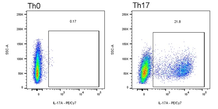
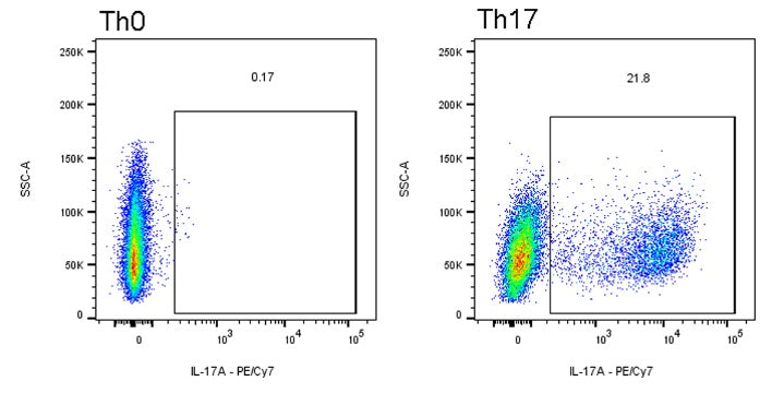
Negative Control Group (Th0) Setup
To evaluate the specificity of polarization, set up a Th0 negative control group in parallel. The procedure is identical to the Th1 induction group described above, except for the use of a different culture medium.
1. Th0 Maintenance Medium:RPMI-1640 supplemented with 10% FBS, 55 μM β-mercaptoethanol, 10 μg/mL anti-human IL-4, 10 μg/mL anti-human IFN-γ, 200 U/mL IL-2, and 1 μg/mL anti-human CD28 mAb.
2. Absence of Polarizing Cytokines: This medium does not contain any polarizing cytokines.
3. The negative control group did not require treatment with PMA, Ionomycin, and Brefeldin A for 5 hours.
Except for the above differences, cell seeding, medium change, and density monitoring were the same as those in the Th17 induction group.
Flow Cytometry Detection
1.Cell Harvesting: Discard the culture supernatant following induction. Wash the cells once with phosphate-buffered saline (PBS). Discard the wash supernatant and gently resuspend any adherent cells in a fresh volume of PBS by pipetting.
2.Cell Counting: Count the cells using an automated cell counter and calculate the total cell number. Centrifuge the cell suspension at 300 × g for 5 minutes at 4°C. Discard the supernatant and resuspend the cell pellet at a concentration of 1×10⁷ cells/mL in PBS.
3.Viability Staining: Add 100 µL of the diluted Fixable Viability Dye 452 (Cat# S0D0021) per 1×10⁶ cells. Refer to the product manual for specific dilution instructions. Incubate as recommended by the manufacture.
4.Cell Washing: Wash the cells by adding an excess volume of PBS. Centrifuge at 300 × g for 5 minutes at 4°C and carefully decant the supernatant to remove residual dye.
5.Cell Fixation: Transfer 100 µL of cell suspension (containing 1×10⁶ cells) to each well of a 96-well U-bottom plate. Centrifuge the plate at 300 × g for 5 minutes at 4°C and discard the supernatant by rapid inversion. Fix cells by adding 200 µL of 4% paraformaldehyde (PFA) per well. Incubate for 15 minutes at room temperature, protected from light.
6.Cell Washing: Centrifuge the plate at 300 × g for 5 minutes at 4°C and discard the supernatant. Add 200 µL of PBS to each well to wash. Repeat centrifugation and discard the supernatant to ensure complete removal of residual fixative.
7.Cell Permeabilization: Add 200 µL of BD Cytofix/Cytoperm™ Fixation/Permeabilization Solution to each well. Incubate for 30 minutes at 4°C.
8.Cell Blocking: Add 100 µL of 1× BD Perm/Wash™ Buffer per well. Include an appropriate concentration of Human IgG (e.g., 0.5-1 µg/µL) in this buffer for Fc receptor blocking. Incubate for 10-15 minutes at 4°C. Centrifuge at 300 × g for 5 minutes at 4°C and discard the supernatant. Note: Step 8 can be performed concurrently with Step 7 by adding the blocking agent directly to the permeabilization solution.
9.Antibody Incubation: Add 100 µL of 1× BD Perm/Wash™ Buffer per well, followed by the appropriately diluted PE-Cyanine7-conjugated IL-17A Monoclonal Antibody according to the manufacturer's instructions.
10.Cell Washing: Centrifuge the plate at 300 × g for 5 minutes at 4°C and discard the supernatant to remove unbound antibody. Add 200 µL of PBS to each well for a final wash. Centrifuge again and discard the supernatant. Resuspend the final cell pellet in 100-200 µL of PBS or a dedicated flow cytometry staining buffer for acquisition.
11.Flow Cytometric Acquisition: Acquire samples on a flow cytometer.
Guidelines
Store in separate containers to reduce the number of freeze-thaw cycles.
Picture
Picture
Bioactivity