WB result of TFAM Recombinant Rabbit mAb
Primary antibody: TFAM Recombinant Rabbit mAb at 1/4000 dilution
Primary antibody incubation conditions: overnight at 4°C
Lane 1: HeLa whole cell lysate 20 µg
Lane 2: Hep G2 whole cell lysate 20 µg
Lane 3: 293T whole cell lysate 20 µg
Lane 4: A-431 whole cell lysate 20 µg
Lane 5: HL-60 whole cell lysate 20 µg
Lane 6: MCF7 whole cell lysate 20 µg
Secondary antibody: Goat Anti-rabbit IgG (H+L), HRP conjugated at 1/10000 dilution
Predicted MW: 29 kDa
Observed MW: 23 kDa
TFAM Recombinant Rabbit mAb
TFAM Recombinant Rabbit mAb
Price:
Regular price
$100 USD
Regular price
Sale price
$100 USD
Unit price
per
For shipping services or bulk orders, you may request a quotation.
Secure checkout with

View full details
Product Details
Product Details
Product Specification
| Host | Rabbit |
| Antigen | TFAM |
| Location | Mitochondrion |
| Accession | Q00059 |
| Antibody Type | Recombinant mAb |
| Isotype | IgG |
| Application | WB, IHC-P, IF |
| Reactivity | Hu, Ms, Rt |
| Positive Sample | HeLa, Hep G2, 293T, A-431, HL-60, MCF7, C2C12, EL4, A20, Neuro-2a, mouse heart, mouse kidney, rat heart, rat kidney |
| Purification | Protein A |
| Concentration | 1.1 mg/ml |
| Conjugation | Unconjugated |
| Physical Appearance | Liquid |
| Storage Buffer | PBS, 40% Glycerol, 0.05% BSA, 0.02% sodium azide |
| Stability & Storage | 12 months from date of receipt / reconstitution, -20 °C as supplied |
Dilution
| application | dilution | species |
| WB | 1:4000-1:20000 | Hu, Ms, Rt |
| IHC-P | 1:2000-1:8000 | Hu, Ms, Rt |
| IF | 1:500 | Hu |
Background
TFAM (mitochondrial transcription factor A, also known as mtTFA/TCF6) is a high-mobility group (HMG) protein encoded by a nuclear gene and transported into the mitochondrial matrix with the assistance of the HSP60-HSP70 chaperone complex. The human protein is approximately 246 amino acids in length and contains two conserved HMG domains that can intercalate between DNA base pairs, inducing an approximately 180° U-shaped bend in the mtDNA promoter region and recognizing sequence features such as GN10G, thereby combining sequence-specific transcriptional regulation with non-sequence-specific DNA packaging functions. As a core scaffold protein of the mitochondrial transcription initiation complex, TFAM cooperates with POLRMT and TFB2M to recruit RNA polymerase upstream of the heavy-strand promoter HSP1 and the light-strand promoter LSP, facilitating promoter recognition, DNA melting, and transcription initiation, and gradually dissociating from the initiation complex after short RNA synthesis. Meanwhile, TFAM wraps and compacts mtDNA in a histone-like manner, maintaining mitochondrial nucleoid structure, mtDNA copy number, replication, and stability. Its expression is regulated by the PGC-1α/NRF axis, p53, FoxM1, and various miRNAs, while its protein stability is controlled by the mitochondrial protease LONP1 and Trim21-mediated ubiquitin-proteasome degradation, with its activity further modulated by PKA phosphorylation and SIRT3 deacetylation. Recent studies have also revealed that TFAM contains an LIR motif, enabling it to bind cytosolic mtDNA under oxidative or inflammatory stress and undergo LC3-mediated autophagic clearance, thereby suppressing excessive activation of the cGAS-STING pathway and inflammatory responses. TFAM dysfunction is associated with mtDNA depletion, oxidative phosphorylation defects, neurodegenerative diseases, cardiovascular diseases, and various cancers, often exhibiting context-dependent dual roles in tumors, making it an important protein marker for assessing mitochondrial function, mtDNA status, and metabolic reprogramming.
Picture
Picture
Western Blot
WB result of TFAM Recombinant Rabbit mAb
Primary antibody: TFAM Recombinant Rabbit mAb at 1/4000 dilution
Primary antibody incubation conditions: overnight at 4°C
Lane 1: C2C12 whole cell lysate 20 µg
Lane 2: EL4 whole cell lysate 20 µg
Lane 3: Neuro-2a whole cell lysate 20 µg
Lane 4: A20 whole cell lysate 20 µg
Lane 5: mouse heart lysate 20 µg
Lane 6: mouse kidney lysate 20 µg
Secondary antibody: Goat Anti-rabbit IgG (H+L), HRP conjugated at 1/10000 dilution
Predicted MW: 29 kDa
Observed MW: 23 kDa
WB result of TFAM Recombinant Rabbit mAb
Primary antibody: TFAM Recombinant Rabbit mAb at 1/4000 dilution
Primary antibody incubation conditions: overnight at 4°C
Lane 1: rat heart lysate 20 µg
Lane 2: rat kidney lysate 20 µg
Secondary antibody: Goat Anti-rabbit IgG (H+L), HRP conjugated at 1/10000 dilution
Predicted MW: 29 kDa
Observed MW: 23-25 kDa
Immunohistochemistry
IHC shows positive staining in paraffin-embedded human colon. Anti-TFAM antibody was used at 1/5000 dilution, followed by a HRP Polymer for Rabbit IgG (ready to use). Counterstained with hematoxylin. Heat mediated antigen retrieval with Tris/EDTA buffer pH9.0 was performed before commencing with IHC staining protocol.
IHC shows positive staining in paraffin-embedded human kidney. Anti-TFAM antibody was used at 1/5000 dilution, followed by a HRP Polymer for Rabbit IgG (ready to use). Counterstained with hematoxylin. Heat mediated antigen retrieval with Tris/EDTA buffer pH9.0 was performed before commencing with IHC staining protocol.
IHC shows positive staining in paraffin-embedded human spleen. Anti-TFAM antibody was used at 1/5000 dilution, followed by a HRP Polymer for Rabbit IgG (ready to use). Counterstained with hematoxylin. Heat mediated antigen retrieval with Tris/EDTA buffer pH9.0 was performed before commencing with IHC staining protocol.
IHC shows positive staining in paraffin-embedded human colon cancer. Anti-TFAM antibody was used at 1/5000 dilution, followed by a HRP Polymer for Rabbit IgG (ready to use). Counterstained with hematoxylin. Heat mediated antigen retrieval with Tris/EDTA buffer pH9.0 was performed before commencing with IHC staining protocol.
IHC shows positive staining in paraffin-embedded human pancreatic cancer. Anti-TFAM antibody was used at 1/5000 dilution, followed by a HRP Polymer for Rabbit IgG (ready to use). Counterstained with hematoxylin. Heat mediated antigen retrieval with Tris/EDTA buffer pH9.0 was performed before commencing with IHC staining protocol.
IHC shows positive staining in paraffin-embedded mouse kidney. Anti-TFAM antibody was used at 1/5000 dilution, followed by a HRP Polymer for Rabbit IgG (ready to use). Counterstained with hematoxylin. Heat mediated antigen retrieval with Tris/EDTA buffer pH9.0 was performed before commencing with IHC staining protocol.
Immunofluorescence
IF shows positive staining in paraffin-embedded human brain. Anti-TFAM antibody was used at 1/500 dilution (Green) and incubated overnight at 4°C. Goat polyclonal Antibody to Rabbit IgG - H&L (Alexa Fluor® 488) was used as secondary antibody at 1/1000 dilution. Counterstained with DAPI (Blue). Heat mediated antigen retrieval with EDTA buffer pH9.0 was performed before commencing with IF staining protocol.
IF shows positive staining in paraffin-embedded human prostate. Anti-TFAM antibody was used at 1/500 dilution (Green) and incubated overnight at 4°C. Goat polyclonal Antibody to Rabbit IgG - H&L (Alexa Fluor® 488) was used as secondary antibody at 1/1000 dilution. Counterstained with DAPI (Blue). Heat mediated antigen retrieval with EDTA buffer pH9.0 was performed before commencing with IF staining protocol.
IF shows positive staining in paraffin-embedded human stomach. Anti-TFAM antibody was used at 1/500 dilution (Green) and incubated overnight at 4°C. Goat polyclonal Antibody to Rabbit IgG - H&L (Alexa Fluor® 488) was used as secondary antibody at 1/1000 dilution. Counterstained with DAPI (Blue). Heat mediated antigen retrieval with EDTA buffer pH9.0 was performed before commencing with IF staining protocol.
IF shows positive staining in paraffin-embedded human tonsil. Anti-TFAM antibody was used at 1/500 dilution (Green) and incubated overnight at 4°C. Goat polyclonal Antibody to Rabbit IgG - H&L (Alexa Fluor® 488) was used as secondary antibody at 1/1000 dilution. Counterstained with DAPI (Blue). Heat mediated antigen retrieval with EDTA buffer pH9.0 was performed before commencing with IF staining protocol.
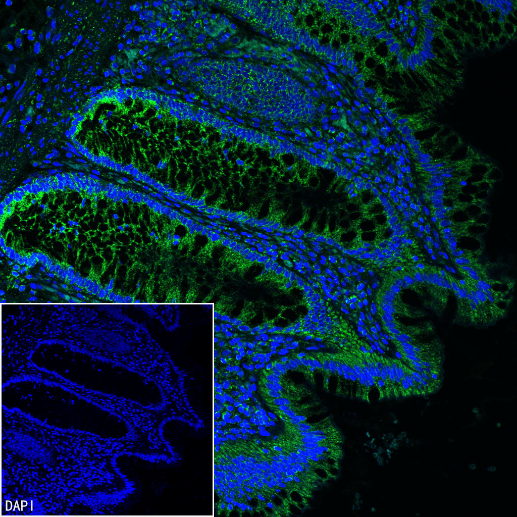
IF shows positive staining in paraffin-embedded human colon. Anti-TFAM antibody was used at 1/500 dilution (Green) and incubated overnight at 4°C. Goat polyclonal Antibody to Rabbit IgG - H&L (Alexa Fluor® 488) was used as secondary antibody at 1/1000 dilution. Counterstained with DAPI (Blue). Heat mediated antigen retrieval with EDTA buffer pH9.0 was performed before commencing with IF staining protocol.