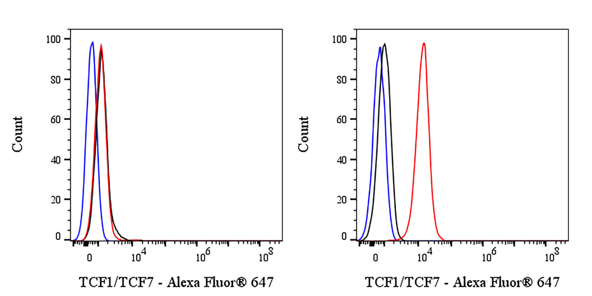
Flow cytometric analysis of 4% PFA fixed 90% methanol permeabilized A20 (Mouse reticulum sarcoma B lymphocyte, left) / EL-4 (Mouse lymphoma T lymphocyte, right) labelling TCF1/TCF7 (Alexa Fluor® 647 Conjugate) antibody at 1/2000 dilution (0.1 μg) / (Red) compared with a Rabbit monoclonal IgG (Black) isotype control and an unlabelled control (cells without incubation with primary antibody and secondary antibody) (Blue). Flow cytometry and data analysis were performed using Agilent NovoCyte Quanteon and FlowJo™ software.
Negative control: A20
TCF1/TCF7 Recombinant Rabbit mAb (Alexa Fluor® 647 Conjugate) (S-3486)
TCF1/TCF7 Recombinant Rabbit mAb (Alexa Fluor® 647 Conjugate) (S-3486)
Price:
Regular price
$176 USD
Regular price
Sale price
$176 USD
Unit price
per
For shipping services or bulk orders, you may request a quotation.
Secure checkout with

View full details
Product Details
Product Details
Product Specification
| Location | Nucleus |
| Accession | P36402 |
| Clone Number | S-3486 |
| Antibody Type | Recombinant mAb |
| Isotype | IgG |
| Application | ICC, ICFCM |
| Reactivity | Hu, Ms |
| Purification | Protein A |
| Concentration | 2 mg/ml |
| Conjugation | Alexa Fluor® 647 |
| Physical Appearance | Liquid |
| Storage Buffer | PBS, 1% BSA, 0.3% Proclin 300 |
| Stability & Storage | 12 months from date of receipt / reconstitution, 2 to 8 °C as supplied |
Dilution
| application | dilution | species |
| ICC | 1:2000 | Hu |
| ICFCM | 1:2000 | Hu, Ms |
Background
TCF1 (T-cell factor 1), encoded by the TCF7 gene, is a 48–58 kDa sequence-specific HMG-box transcription factor and a founding member of the TCF/LEF family that serves as the primary nuclear effector of the canonical Wnt/β-catenin pathway, binding via its N-terminal β-catenin interaction domain and its central DNA-binding HMG motif to the consensus sequence 5’-(A/T)(A/T)CAAAG-3’ in promoters/enhancers of genes such as MYC, CCND1 and LEF1, thereby driving embryonic stem cell pluripotency, thymic T-cell lineage specification, CD8⁺ T-cell memory formation, and adult intestinal epithelial renewal, while its expression is dynamically regulated by alternative promoters and differential splicing to yield long (p45) and short (p33) isoforms with opposing activities; TCF1 also possesses context-dependent repressive functions when β-catenin is absent, interacts with Groucho/TLE co-repressors, and is frequently inactivated by loss-of-function mutations or deletions in colorectal, hematopoietic and other malignancies, making it both a developmental gatekeeper and a tumor suppressor.
Picture
Picture
FC
Immunocytochemistry
ICC shows positive staining in EL-4 cells (top panel) and negative staining in A20 cells (below panel). Anti-TCF1/TCF7 (Alexa Fluor® 647 Conjugate) antibody was used at 1/2000 dilution (magenta) and incubated overnight at 4°C. The cells were fixed with 4% PFA and permeabilized with 0.1% PBS-Triton X-100. Nuclei were counterstained with DAPI (Blue).