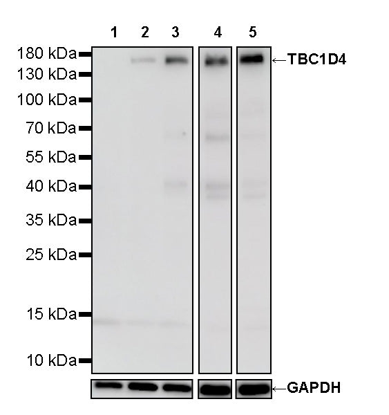
WB result of TBC1D4 Recombinant Rabbit mAb
Primary antibody: TBC1D4 Recombinant Rabbit mAb at 1/1000 dilution
Lane 1: A431 whole cell lysate 20 µg
Lane 2: HeLa whole cell lysate 20 µg
Lane 3: HepG2 whole cell lysate 20 µg
Lane 4: HCT 116 whole cell lysate 20 µg
Lane 5: Jurkat whole cell lysate 20 µg
Low expression control: A431 whole cell lysate 20 µg
Secondary antibody: Goat Anti- rabbit IgG, (H+L), HRP conjugated at 1/10000 dilution
Predicted MW: 147 kDa
Observed MW: 160 kDa
This blot was developed with high sensitivity substrate.
TBC1D4 Recombinant Rabbit mAb (S-4328-80)
TBC1D4 Recombinant Rabbit mAb (S-4328-80)
Price:
Regular price
$100 USD
Regular price
Sale price
$100 USD
Unit price
per
For shipping services or bulk orders, you may request a quotation.
Secure checkout with

View full details
Product Details
Product Details
Product Specification
| Host | Rabbit |
| Antigen | TBC1D4 |
| Synonyms | TBC1 domain family member 4; AS160; KIAA0603 |
| Immunogen | Recombinant Protein |
| Location | Cytoplasm |
| Accession | O60343 |
| Clone Number | S-4328-80 |
| Antibody Type | Recombinant mAb |
| Isotype | IgG |
| Application | WB |
| Reactivity | Hu |
| Positive Sample | HeLa, HepG2, HCT 116, Jurkat |
| Purification | Protein A |
| Concentration | 2 mg/ml |
| Conjugation | Unconjugated |
| Physical Appearance | Liquid |
| Storage Buffer | PBS, 40% Glycerol, 0.05% BSA, 0.02% sodium azide |
| Stability & Storage | 12 months from date of receipt / reconstitution, -20 °C as supplied |
Dilution
| application | dilution | species |
| WB | 1:1000 | Hu |
Background
TBC1D4, also known as AS160 (Akt substrate of 160 kDa), is a member of the Tre-2/BUB2/CDC16 (TBC) domain family and plays a critical role in regulating glucose metabolism in insulin-target tissues such as skeletal muscle and adipose tissue. This protein functions essentially as a Rab-GTPase-activating protein (RabGAP) and possesses a refined structure, containing two N-terminal phosphotyrosine-binding domains (PTB1 and PTB2), a central calmodulin-binding domain, a C-terminal catalytic RabGAP domain, and multiple motifs that can be phosphorylated by protein kinase B (AKT). Under basal conditions without insulin stimulation, TBC1D4 acts through its GAP domain on Rab proteins (such as Rab2A, Rab8A, Rab10, and Rab14), converting them into the inactive GDP-bound state, thereby anchoring glucose transporter 4 (GLUT4) within intracellular vesicles and preventing its translocation to the plasma membrane. Upon insulin signal activation, activated AKT phosphorylates multiple critical sites on TBC1D4, including Thr642. This phosphorylation event both promotes the dissociation of TBC1D4 from GLUT4 vesicles and facilitates the binding of 14-3-3 proteins, effectively inhibiting its RabGAP activity. Consequently, Rab proteins are maintained in their active state, driving GLUT4-containing vesicles to translocate and fuse with the plasma membrane, thereby efficiently facilitating cellular glucose uptake from the bloodstream. Furthermore, studies have revealed that TBC1D4 can form homodimers via its C-terminal region, and homozygous mutations in this gene significantly increase an individual's risk of developing type 2 diabetes, characterized by higher levels of both blood glucose and insulin following glucose ingestion.
Picture
Picture
Western Blot