Human T Cell Activation Kit (Magnetic Bead )
Human T Cell Activation Kit (Magnetic Bead )

Product Details
Product Details
Product Specification
| Storage Buffer | |
||||
| Reconstitution | IL-2: Reconstitute at 0.1-1 mg/ml according to the size in ultrapure water after rapid centrifugation. |
||||
| Stability & Storage |
|
Components
Contain |
Formulation |
Reference dosage |
For 10^8cells |
For 4*10^8cells |
|
IL-2 Protein, Human |
Lyophilized Powder |
10 ng/mL |
5μg |
10μg |
|
T Cell Activation Nanobeads,Human(RUO)
|
Liquid |
5 μL/1×106cells |
0.5mL |
2mL |
Protocol
Complete Medium Formulation for Reference:
Component |
Final Concentration |
RPMI-1640 |
90% |
Fetal Bovine Serum |
10% |
Sodium Pyruvate 100× |
1× |
L-Glutamine 100× |
1× |
HEPES |
1× |
Penicillin-Streptomycin |
1× |
β-Mercaptoethanol |
55 μM |
Procedure
1. Prepare a single‑cell suspension of human peripheral blood mononuclear cells (PBMCs). Perform cell counting using AO/PI to determine the cell number required. Resuspend 1×10⁶ cells in 995 μL of complete T‑cell medium. For T‑cell activation, the recommended concentration is (1–3)×10⁶ cells/mL. For a 24‑well plate, a concentration of 1×10⁶ cells/mL is recommended.
2. Add the following cytokines and beads to the cell suspension from step 1:
- Add 5 μL of T Cell Activation Nanobeads per 1×10⁶ cells and mix thoroughly but gently.
- Add 10 ng/mL human interleukin‑2 (IL‑2) protein.
3. Transfer the cells to a 24‑well plate and incubate statically in a 37 °C, 5 % CO₂ incubator for 48 h.
4. After 48 h of culture, check cell health and record images.
5. Centrifuge the cell suspension at 400×g for 5 minutes and discard the supernatant. Wash the cell pellet with PBS.
6. Centrifuge the cell suspension at 400×g for 5 minutes and discard the supernatant. Resuspend the cells in 0.5 mL of 1 % bovine serum albumin (BSA) and count the cells. Assess cell viability, which should be >95 %.
7. Block 1×10⁶ cells with 10 µg of human IgG antibody. Incubate on ice for 30 minutes.
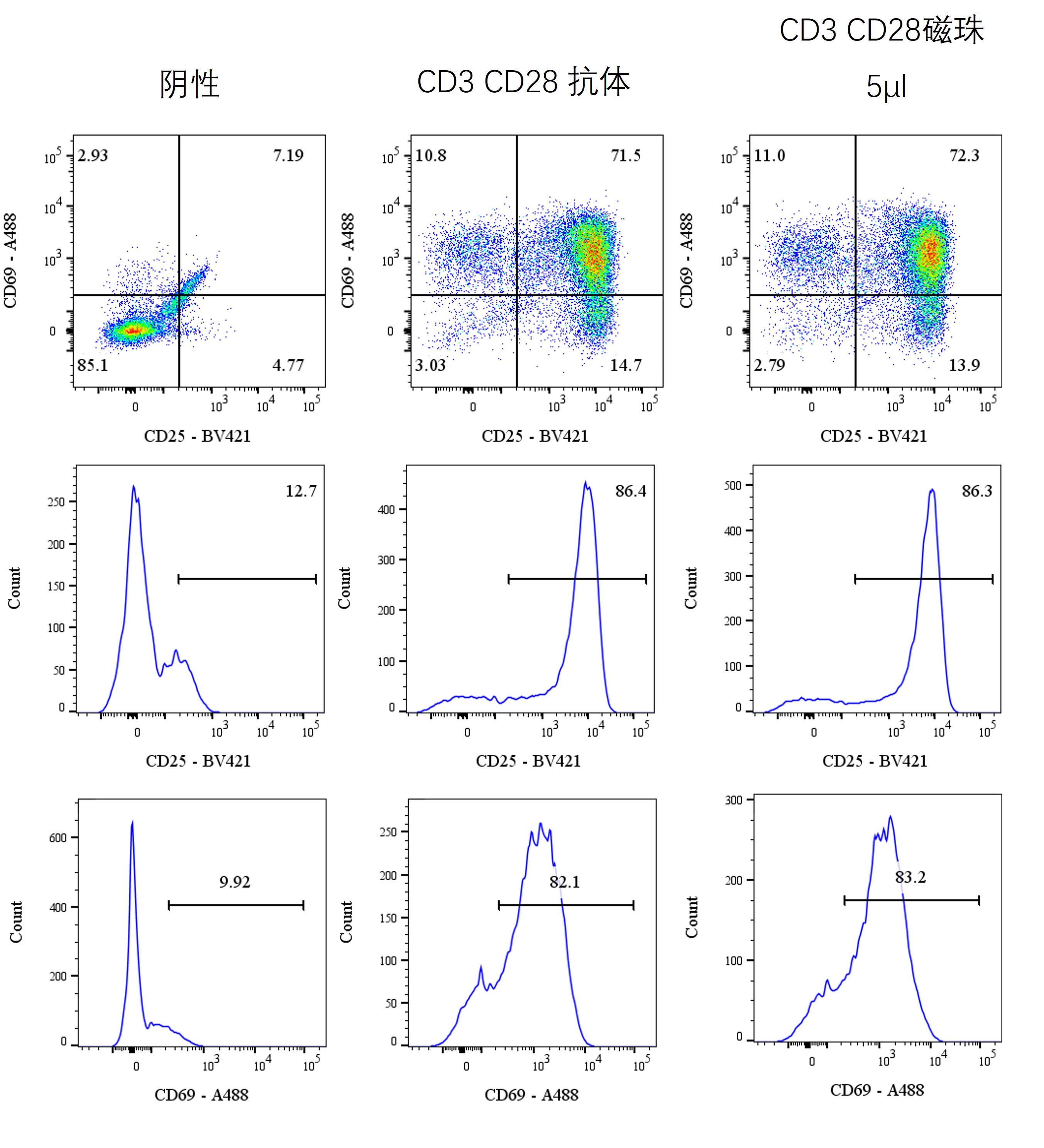
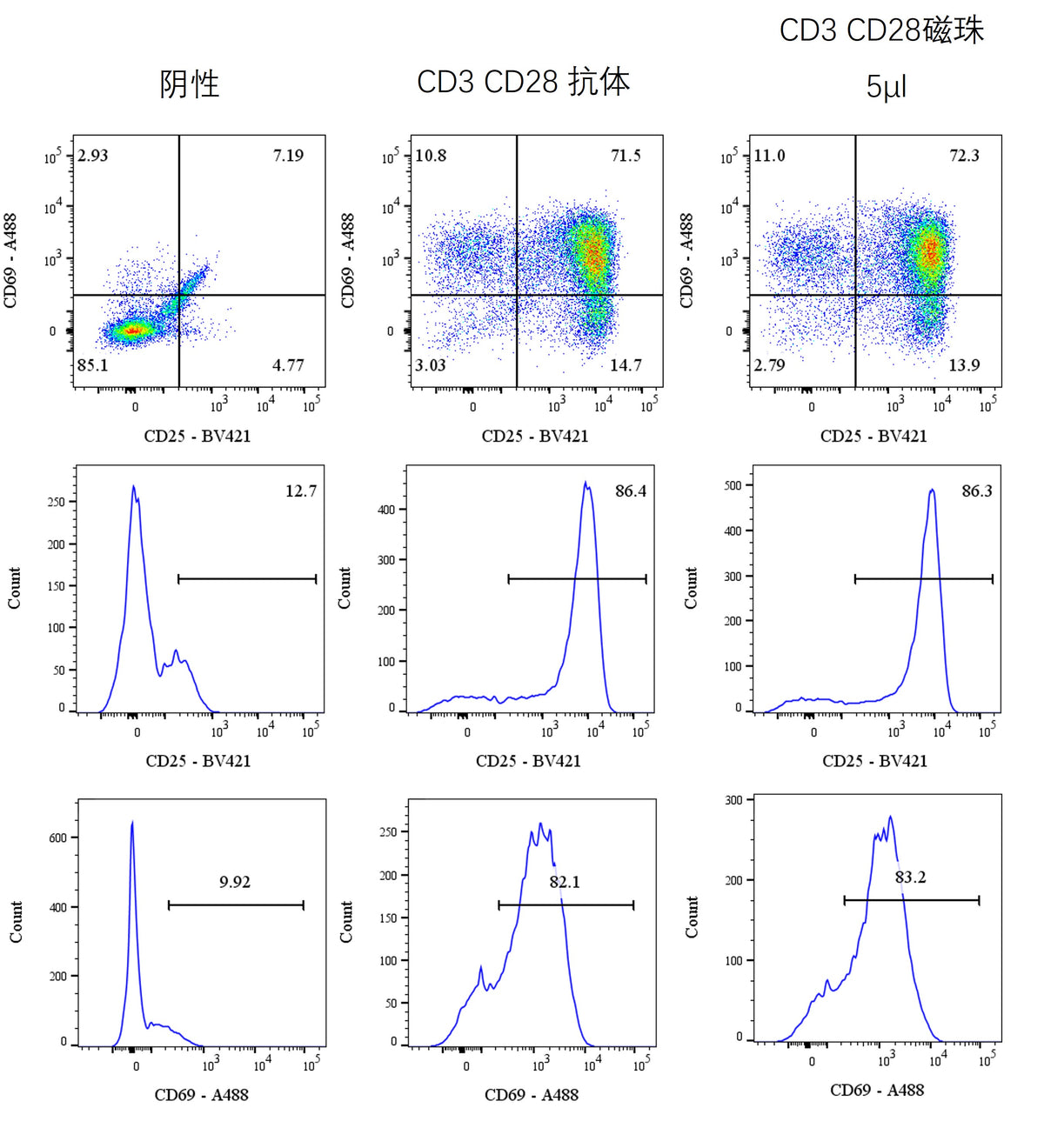
8. According to the manufacturer’s instructions, add Brilliant Violet 421™ anti‑human CD25 antibody and Alexa Fluor® 488 anti‑human CD69 antibody, and incubate at 4 °C for 30 minutes.
9. Centrifuge the cell suspension at 400×g for 5 minutes and wash the cell pellet twice with PBS containing 1 % BSA.
10. Resuspend the cell pellet in 200 µL of PBS for flow cytometric analysis (≥10,000 cells are required).
Guidelines
1. T Cell Activation Nanobeads, Human (RUO) should be thoroughly mixed before use. During the operation, efforts should be made to minimize bubble formation to avoid any impact on subsequent operations.
2. All operations throughout the process should be conducted in a sterile environment.
3. For the initial activation, it is necessary to ensure that the reagents and cells remain in contact for 2 to 3 days. Removing the reagents too early will result in insufficient T cell activation and decreased proliferation ability.
4. T cells can undergo virus transduction experiments 2 days after activation. It is necessary to independently explore the optimal transduction titer and timing.
5. The residual EDTA in the cell system will significantly inhibit T cell activation stimulation. Before the experiment, it is necessary to thoroughly remove it.
Picture
Picture
FC