Mouse spleen cells were sorted to obtain mouse T cells, CD4 T cells, and CD8 T cells. These cells were stimulated and activated using the T Cell Activation/Expansion Kit for mice, and after 2 days of culture, they were stained with antibodies targeting the activation markers CD25-Alexa Fluor647 (SDT Cat: S0B1545) and CD69-Alexa Fluor488 (SDT Cat: S0B8295). Flow cytometry results showed that the positive expression rates of the T-cell activation markers were significantly increased following magnetic bead-mediated activation, indicating a high level of cell activation.
T Cell Activation/Expansion Kit, mouse(RUO)
T Cell Activation/Expansion Kit, mouse(RUO)
Price:
Regular price
$420 USD
Regular price
Sale price
$420 USD
Unit price
per
For shipping services or bulk orders, you may request a quotation.
Secure checkout with

View full details
Product Details
Product Details
Product Specification
| Format |
2 mL Streptavidin NanoBeads, in a PBS buffer containing BSA and Poloxamer 188; 0.4 mL CD3ε-Biotin, mouse (100 µg/mL), in a PBS buffer containing BSA; 0.4 mL CD28-Biotin, mouse (100 µg/mL), in a PBS buffer containing BSA; |
| Capacity | Can activate 2 × 10⁸ T cells; |
| Principle of Separation |
This kit is used for the in vitro activation and expansion of mouse T cells, with its core components consisting of streptavidin magnetic beads and biotin‑labeled mouse CD3ε/CD28 antibodies. After biotinylated CD3ε and CD28 antibodies are conjugated to the surface of streptavidin‑nanomagnetic beads, the beads can mimic antigen‑presenting cells, stimulating the activation of single‑cell suspensions from the spleen or lymph nodes, or purified resting T cells; on days 6–8 of culture, they can be restimulated to achieve large‑scale T‑cell expansion. |
| Applications in cell sorting | This product is intended for research use only and is not to be used for clinical diagnosis or treatment. |
| Reactivity | Mouse |
| Magnetic Bead Size | 150 nm |
| Stability & Storage | Store protected from light at 2–8 °C; do not freeze. |
Background
MouseTLymphocytes are the core effector cells of the adaptive immune system, mainly divided intoCD4+helperTcells andCD8+cytotoxicTcells, playing a key role in anti-infectious immunity, tumor immunity, immune regulation, and the development of autoimmune diseases. Under physiological conditions, naïveTcells in the mouse spleen and lymph nodes are in a quiescent, dormant state, barelyproliferating,expressing low levels of functional molecules, and thus unable to directly conduct in vitro functional experiments. Therefore, controlledTcell activation and large-scale expansion in vitro are core foundational technologies for studying murine immune mechanisms, validating cell functions, and constructing immune models; this reagentbox employsartificially simulated antigen-presenting cell technology, using biotinylatedCD3εandCD28antibody-conjugated anti-biotin magnetic beads to precisely replicateTcell activation's dual signals in vitro, achieving highly efficient, non-specific activation and expansion of polyclonalTcells, suitable for total mouseTcells,CD4T,CD8 TMass amplification of cells. Magneticbeads serve assolid-phase carriers, capable of simultaneously enriching large quantities ofCD3εandCD28antibodies, stably binding to the corresponding receptors on the surface ofTcells, continuously and evenly providing the first and second signals, thereby avoiding the instability inherent in natural antigen presentation and addressingprimary mouseTcell issues of poor in vitro activation and weak proliferation specific to certain species, significantly enhancing activation efficiency and expansion multiples.
Protocol
1. Experimental reagents
1.1 Buffer:pH 7.2 PBSbuffer, with the addition of2 mM EDTA,2–8℃Pre-cooled; do not use buffers containing calcium or magnesium ions/Culture medium;
1.2 Cell culture medium: RPMI 1640, with the addition of10%fetal bovine serum (FBS)+IL-2(final concentration30–100 IU/mL);Optional addition of50-100μMβ-mercaptoethanol, to maintain cell viability under rapid proliferation conditions;
2. Magnetic bead conjugation (conjugatingCD3ε/CD28antibodies)
The magnetic beads contain no preservatives; all aliquoting operations must be performed under sterile conditions; it is recommended to conjugate500 μLof magnetic beads at a time, and after conjugation, the beads2–8℃can be stably stored for3months without washing away unbound antibodies.
2.1 Take2 mLof a sterile centrifuge tube, and add100μLCD3ε-Biotin + 100μLCD28-Biotin, mix thoroughly by pipetting;
2.2 add300μL buffer (pH 7.2 PBScontaining2 mM EDTA) and mix well;
2.3 thoroughly vortexto resuspend the magnetic beads, ensuring uniform dispersion of the beads; then pipette500μLStreptavidinNanoBeadsadd to the above antibody mixture, bringing the total volumeto 1 mL;
2.4 place the centrifuge tube on a rotator,at 2–8℃and incubate with gentle continuous rotationfor 1 hour; this results in coupledCD3ε/CD28magnetic beads, which can be used directly afterward;
3. Experimental steps
All operations must be performed in a sterile environment.
3.1 Sample preparation
Resting mouse T cells derived from the spleenTCell activation and expansion, preparing single-cell suspensions from lymphoid organs (spleen, lymph nodes). Magnetic bead sorting can be used.TCell activation and expansion: positive selectionCD3/CD4/CD8 T cells:CD3ε Nanobeads Kit, mouse (RUO)(Cat. No. S0K0015);CD4 Nanobeads,Mouse(RUO) (Cat. No. S0K0003);CD8 Nanobeads, Mouse(RUO)(Cat. No. S0K0005);negative selec-tionCD3/ CD4/CD8T cells::Mouse T Cell Isolation Kit(Cat. No. S0K1008/S0K3006);Mouse CD4 T Cell Isolation Kit(Cat. No. S0K1005/S0K3003); Mouse CD8 T Cell Isolation Kit (Cat. No. S0K1007/S0K3004).
Refer to the corresponding product manual for specific operations.
3.2TCell activation and expansion procedures
The basic system in this protocol is suitable for1×10⁶cellsafter sortingTcellscorresponding to20μLof coupled magnetic beads; when the cell quantity doubles, all reagent volumes are scaled up accordingly.
3.2.1 Take the sorted cells at a concentration of1×10⁶/mLand resuspend them infresh complete culture medium (RPMI1640 + 10% FBS + 30–100 IU/mL IL-2)in,take1mLof cellsinto a centrifuge tube,thoroughly mix the coupled magnetic beads, then add20μLof the coupledmagnetic beads,centrifuge at400 gfor7 min;(negative control: use uncoupled blank magnetic beads instead ofcoupledmagnetic beads;)
3.2.2 After centrifugation, discard the supernatant and resuspend the pellet in1 mLFresh complete medium (RPMI1640 + 10% FBS + 30–100 IU/mL IL-2),Add the cells to the corresponding plates for culture. To ensure optimal stimulation and proliferation, it is recommended to use a standard seeding density:1×10⁶cells/cm²;
Optimal Culture System Ratio Table for Each Culture Plate
Culture Plate Model |
Growth Base Area |
Total CultureVolume |
TCell Seeding Quantity |
Amount of Coupled Magnetic Beads |
96Well Plate |
0.31 cm² |
0.2 mL |
0.25×10⁶ |
5 μL |
48Well Plate |
1 cm² |
1 mL |
1×10⁶ |
20μL |
24Well Plate |
2 cm² |
2 mL |
2×10⁶ |
40μL |
12Well Plate |
4 cm² |
4 mL |
4×10⁶ |
80μL |
6Well Plate |
10 cm² |
5 mL |
5×10⁶ |
100μL |
3.2.3 Place the culture plate ina CO₂incubator for cultivation, observe cell status daily, and replenish the medium promptly when it becomes insufficient;
3.2.4 After culturing for2days, gently disperse cell clumps; at this step, samples can be taken for flow cytometry analysis.mouse CD25, mouse CD69to assess cell activation status.;
3.2.5 Passage and expansion: every2days, subculture at a ratio of1:2or1:4proportion, add fresh culture medium, and if proliferation is too rapid, shorten the passage interval.;
3.2.6 Secondary stimulation: after activating the culture for6–8days,Tcells enter a quiescentstate,if continued expansion is needed, magnetic beads must be added again to restimulate.
3.3 Magnetic bead removal procedure
If downstream operations require magnetic bead sorting, replacement of stimulating antigens, or stimulants, the magnetic beads must be removed in advance:
3.3.1 Collect the cells and transfer them to5/15/50 mLCentrifuge the tube and wash once with buffer;
3.3.2 Resuspend the cells in buffer, at a maximum concentration2×10⁷cells/mL, and pipette thoroughly to mix evenly;
3.3.3 Fix the centrifuge tube on the magnetic separator and let it stand for adsorption10 min;
3.3.4 Transfer the cells to a new centrifuge tube, collect the bead-free cells, and proceed with subsequent experiments.
Picture
Picture
Validation Data
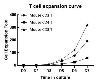
Mouse spleen cells were sorted to obtain Mouse T, CD4 T, and CD8 T cells, which were then activated using the T Cell Activation/Expansion kit for mice and cultured for 7 days.
T cell complete culture medium: RPMI 1640 basal medium supplemented with 10% FBS, 50 IU/mL IL-2, and 50 μM β‑mercaptoethanol.
Seeding density: 0.5 × 10⁶ cells/mL.
Cell expansion curve on day 7 of culture.
DLS
·