WB result of SV40 T-antigen Recombinant Mouse mAb
Primary antibody: SV40 T-antigen Recombinant Mouse mAb at 1/1000 dilution
Lane 1: HUVEC whole cell lysate 20 µg
Lane 2: HEK-293T whole cell lysate 20 µg
Secondary antibody: Goat Anti-Mouse IgG (H+L), HRP conjugated at 1/10000 dilution
Predicted MW: 90 kDa
Observed MW: 90kDa
SV40 T-antigen Recombinant Mouse mAb (S-6032)
SV40 T-antigen Recombinant Mouse mAb (S-6032)
Price:
Regular price
$100 USD
Regular price
Sale price
$100 USD
Unit price
per
For shipping services or bulk orders, you may request a quotation.
Secure checkout with

View full details
Product Details
Product Details
Product Specification
| Host | Mouse |
| Antigen | SV40 T-antigen |
| Clone Number | S-6032 |
| Antibody Type | Mouse mAb |
| Isotype | IgG2a |
| Application | WB, ICC |
| Reactivity | Hu, Mk |
| Positive Sample | HUVEC, HEK-293T, COS-7 |
| Purification | Protein A |
| Concentration | 2 mg/ml |
| Conjugation | Unconjugated |
| Physical Appearance | Liquid |
| Storage Buffer | PBS, 40% Glycerol, 0.05% BSA, 0.02% sodium azide |
| Stability & Storage | 12 months from date of receipt / reconstitution, -20 °C as supplied |
Dilution
| application | dilution | species |
| WB | 1:0000 | Hu, Mk |
| ICC | 1:500 | Hu |
Background
SV40 large T-antigen is a multifunctional nuclear phosphoprotein encoded by the early region of simian virus 40 (SV40), with a molecular weight of approximately 90–100 kDa. It is both an essential factor for viral DNA replication and capsid assembly in permissive cells and the principal oncoprotein responsible for cell transformation and tumorigenesis by this virus in non-permissive cells. Its functions are highly modular: the amino-terminal region binds to and inactivates the host cell's key tumor suppressors pRB and p53, relieving cell cycle arrest and inhibiting apoptosis, thereby driving cells into S phase and creating a favorable environment for viral replication; the central domain confers sequence-specific DNA-binding ability and helicase/ATPase activity, enabling it to recognize the viral replication origin on its own and initiate DNA replication—a property that has made SV40 a classic model for studying the mechanisms of eukaryotic DNA replication initiation. T-antigen also contains a nuclear localization signal that allows it to enter the nucleus to carry out replication and transcriptional regulation functions, while a small fraction of T-antigen can also localize to the plasma membrane, and these two subcellular localization forms may act synergistically to mediate the full transformation phenotype. Because a single viral protein is sufficient to initiate complete DNA replication, SV40 large T-antigen has for decades been a powerful experimental tool for dissecting cell cycle regulation, tumor suppressor pathways, and eukaryotic replication mechanisms.
Picture
Picture
Western Blot
WB result of SV40 T-antigen Recombinant Mouse mAb
Primary antibody: SV40 T-antigen Recombinant Mouse mAb at 1/1000 dilution
Lane 1: COS-7 whole cell lysate 20 µg
Secondary antibody: Goat Anti-Mouse IgG (H+L), HRP conjugated at 1/10000 dilution
Predicted MW: 90 kDa
Observed MW: 90kDa
Immunocytochemistry
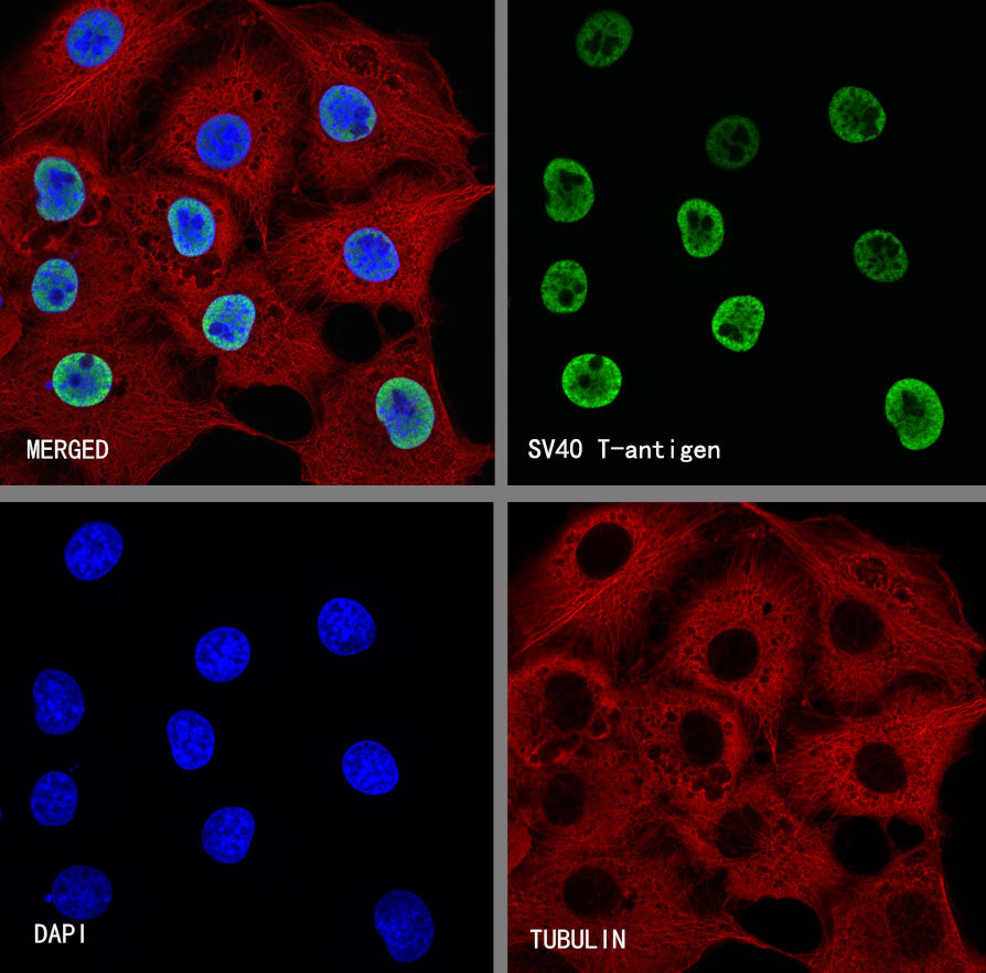
ICC shows positive staining in HUVEC cells. Anti- SV40 T-antigen antibody was used at 1/500 dilution (Green) and incubated overnight at 4°C. Goat polyclonal Antibody to Mouse IgG - H&L (Alexa Fluor® 488) was used as secondary antibody at 1/1000 dilution. The cells were fixed with 4% PFA and permeabilized with 0.1% PBS-Triton X-100. Nuclei were counterstained with DAPI (Blue). Counterstain with tubulin (Red).
ICC shows positive staining in COS-7 cells. Anti- SV40 T-antigen antibody was used at 1/500 dilution (Green) and incubated overnight at 4°C. Goat polyclonal Antibody to Mouse IgG - H&L (Alexa Fluor® 488) was used as secondary antibody at 1/1000 dilution. The cells were fixed with 4% PFA and permeabilized with 0.1% PBS-Triton X-100. Nuclei were counterstained with DAPI (Blue). Counterstain with tubulin (Red).