WB result of STEAP3 Recombinant Rabbit mAb
Primary antibody: STEAP3 Recombinant Rabbit mAb at 1/1000 dilution
Lane 1: Jurkat whole cell lysate 20 µg
Lane 2: 22Rv1 whole cell lysate 20 µg
Lane 3: PC-3 whole cell lysate 20 µg
Lane 4: HepG2 whole cell lysate 20 µg
Negative control: Jurkat whole cell lysate 20 µg
Secondary antibody: Goat Anti-rabbit IgG (H+L), HRP conjugated at 1/10000 dilution
Predicted MW: 55 kDa
Observed MW: 36-58 kDa
STEAP3 Recombinant Rabbit mAb (S-4466-26)
STEAP3 Recombinant Rabbit mAb (S-4466-26)
Price:
Regular price
$100 USD
Regular price
Sale price
$100 USD
Unit price
per
For shipping services or bulk orders, you may request a quotation.
Secure checkout with

View full details
Product Details
Product Details
Product Specification
| Host | Rabbit |
| Antigen | STEAP3 |
| Immunogen | Recombinant Protein |
| Location | Cell membrane |
| Accession | Q658P3 |
| Clone Number | S-4466-26 |
| Antibody Type | Recombinant mAb |
| Isotype | IgG |
| Application | WB, IHC-P, IF |
| Reactivity | Hu, Ms, Rt |
| Positive Sample | 22Rv1, PC-3, HepG2, mouse liver |
| Purification | Protein A |
| Concentration | 0.5 mg/ml |
| Conjugation | Unconjugated |
| Physical Appearance | Liquid |
| Storage Buffer | PBS, 40% Glycerol, 0.05% BSA, 0.02% sodium azide |
| Stability & Storage | 12 months from date of receipt / reconstitution, -20 °C as supplied |
Dilution
| application | dilution | species |
| WB | 1:1000 | Hu, Ms, Rt |
| IHC-P | 1:500-1:2000 | Hu, Ms, Rt |
Background
STEAP3 protein, also known as TSAP6, is an important metalloreductase that plays a central role in cellular iron and copper metabolism. This protein is composed of an N-terminal cytosolic oxidoreductase domain and a C-terminal heme-containing transmembrane domain, and it is primarily localized on endosomal membranes. Its classic function is to act as an endosomal iron reductase during red blood cell development, reducing ferric iron (Fe³⁺) released from transferrin to ferrous iron (Fe²⁺) so that it can be transported into the cytoplasm by divalent metal transporter 1 (DMT1)—a key step in transferrin-dependent iron uptake. STEAP3 also participates in copper homeostasis regulation, capable of reducing cupric copper to cuprous copper. Additionally, STEAP3 is a direct transcriptional target gene of p53, participates in the DNA damage-induced p53-dependent exosome secretion pathway, and plays a role in regulating apoptosis and the cell cycle. In terms of disease associations, mutations in the STEAP3 gene can cause hypochromic microcytic anemia with iron overload (AHMIO2). Recent studies have found that STEAP3 is abnormally expressed in various tumors, such as hepatocellular carcinoma, cervical cancer, renal cell carcinoma, and glioblastoma, and its high expression is generally associated with poor prognosis. In tumor initiation and progression, STEAP3 promotes tumor cell proliferation, migration, and epithelial-mesenchymal transition by regulating the JAK/STAT3 signaling pathway, while also influencing the ferroptosis process through the p53/xCT pathway.
Picture
Picture
Western Blot
WB result of STEAP3 Recombinant Rabbit mAb
Primary antibody: STEAP3 Recombinant Rabbit mAb at 1/1000 dilution
Lane1: HEK-293T cells transfected with a human STEAP2 expression vector containing a myc-his tag
Lane2: HEK-293T cells transfected with a human STEAP3 expression vector containing a myc-his tag
Lane3: HEK-293T cells transfected with a human STEAP4 expression vector containing a myc-his tag
Lane4: HEK-293T cells transfected with a human STEAP1B1 expression vector containing a myc-his tag
Lane5: HEK-293T cells transfected with a human STEAP1B2 expression vector containing a myc-his tag
Lane6: HEK-293T cells transfected with an empty vector containing a myc-his tag
Secondary antibody: Goat Anti-rabbit IgG (H+L), HRP conjugated at 1/10000 dilution
Predicted MW: 55 kDa
Observed MW: 38-60 kDa
WB result of STEAP3 Recombinant Rabbit mAb
Primary antibody: STEAP3 Recombinant Rabbit mAb at 1/1000 dilution
Lane 1: mouse liver lysate 20 µg
Secondary antibody: Goat Anti-rabbit IgG (H+L), HRP conjugated at 1/10000 dilution
Predicted MW: 55 kDa
Observed MW: 38-58 kDa
WB result of STEAP3 Recombinant Rabbit mAb
Primary antibody: STEAP3 Recombinant Rabbit mAb at 1/1000 dilution
Lane 1: rat liver lysate 20 µg
Secondary antibody: Goat Anti-rabbit IgG (H+L), HRP conjugated at 1/10000 dilution
Predicted MW: 55 kDa
Observed MW: 38-58 kDa
Immunohistochemistry
IHC shows positive staining in paraffin-embedded human liver. Anti-STEAP3 antibody was used at 1/2000 dilution, followed by a HRP Polymer for Rabbit IgG (ready to use). Counterstained with hematoxylin. Heat mediated antigen retrieval with Tris/EDTA buffer pH9.0 was performed before commencing with IHC staining protocol.
Negative control: IHC shows negative staining in paraffin-embedded human testis. Anti-STEAP3 antibody was used at 1/2000 dilution, followed by a HRP Polymer for Rabbit IgG (ready to use). Counterstained with hematoxylin. Heat mediated antigen retrieval with Tris/EDTA buffer pH9.0 was performed before commencing with IHC staining protocol.
Negative control: IHC shows negative staining in paraffin-embedded human hepatocellular carcinoma. Anti-STEAP3 antibody was used at 1/2000 dilution, followed by a HRP Polymer for Rabbit IgG (ready to use). Counterstained with hematoxylin. Heat mediated antigen retrieval with Tris/EDTA buffer pH9.0 was performed before commencing with IHC staining protocol.
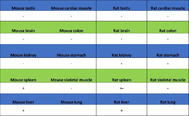
IHC shows positive staining in paraffin-embedded mouse liver. Anti-STEAP3 antibody was used at 1/500 dilution, followed by a HRP Polymer for Rabbit IgG (ready to use). Counterstained with hematoxylin. Heat mediated antigen retrieval with Tris/EDTA buffer pH9.0 was performed before commencing with IHC staining protocol.
Negative control: IHC shows negative staining in paraffin-embedded mouse colon. Anti-STEAP3 antibody was used at 1/500 dilution, followed by a HRP Polymer for Rabbit IgG (ready to use). Counterstained with hematoxylin. Heat mediated antigen retrieval with Tris/EDTA buffer pH9.0 was performed before commencing with IHC staining protocol.
IHC shows positive staining in paraffin-embedded rat liver. Anti-STEAP3 antibody was used at 1/500 dilution, followed by a HRP Polymer for Rabbit IgG (ready to use). Counterstained with hematoxylin. Heat mediated antigen retrieval with Tris/EDTA buffer pH9.0 was performed before commencing with IHC staining protocol.
Negative control: IHC shows negative staining in paraffin-embedded rat testis. Anti-STEAP3 antibody was used at 1/500 dilution, followed by a HRP Polymer for Rabbit IgG (ready to use). Counterstained with hematoxylin. Heat mediated antigen retrieval with Tris/EDTA buffer pH9.0 was performed before commencing with IHC staining protocol.
IHC shows positive staining in paraffin-embedded STEAP3 transfected 293F cells. Anti-STEAP3 antibody was used at 1/8000 dilution, followed by a HRP Polymer for Rabbit IgG (ready to use). Counterstained with hematoxylin. Heat mediated antigen retrieval with Tris/EDTA buffer pH9.0 was performed before commencing with IHC staining protocol.
Negative control: IHC shows negative staining in paraffin-embedded mock transfected 293F cells. Anti-STEAP3 antibody was used at 1/8000 dilution, followed by a HRP Polymer for Rabbit IgG (ready to use). Counterstained with hematoxylin. Heat mediated antigen retrieval with Tris/EDTA buffer pH9.0 was performed before commencing with IHC staining protocol.
Negative control: IHC shows negative staining in paraffin-embedded STEAP1 transfected 293F cells. Anti-STEAP3 antibody was used at 1/8000 dilution, followed by a HRP Polymer for Rabbit IgG (ready to use). Counterstained with hematoxylin. Heat mediated antigen retrieval with Tris/EDTA buffer pH9.0 was performed before commencing with IHC staining protocol.
Negative control: IHC shows negative staining in paraffin-embedded STEAP1B1 transfected 293F cells. Anti-STEAP3 antibody was used at 1/8000 dilution, followed by a HRP Polymer for Rabbit IgG (ready to use). Counterstained with hematoxylin. Heat mediated antigen retrieval with Tris/EDTA buffer pH9.0 was performed before commencing with IHC staining protocol.
Negative control: IHC shows negative staining in paraffin-embedded STEAP1B2 transfected 293F cells. Anti-STEAP3 antibody was used at 1/8000 dilution, followed by a HRP Polymer for Rabbit IgG (ready to use). Counterstained with hematoxylin. Heat mediated antigen retrieval with Tris/EDTA buffer pH9.0 was performed before commencing with IHC staining protocol.
Negative control: IHC shows negative staining in paraffin-embedded STEAP2 transfected 293F cells. Anti-STEAP3 antibody was used at 1/8000 dilution, followed by a HRP Polymer for Rabbit IgG (ready to use). Counterstained with hematoxylin. Heat mediated antigen retrieval with Tris/EDTA buffer pH9.0 was performed before commencing with IHC staining protocol.
Negative control: IHC shows negative staining in paraffin-embedded STEAP4 transfected 293F cells. Anti-STEAP3 antibody was used at 1/8000 dilution, followed by a HRP Polymer for Rabbit IgG (ready to use). Counterstained with hematoxylin. Heat mediated antigen retrieval with Tris/EDTA buffer pH9.0 was performed before commencing with IHC staining protocol.