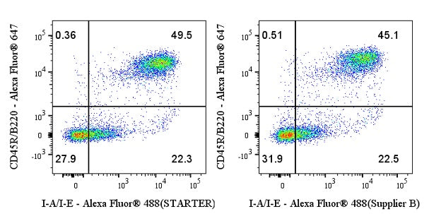
C57BL/6 mouse splenocytes were stained with Alexa Fluor® 647 Rat Anti-Mouse CD45R/B220 Antibody and either Rat Anti-Mouse I-A/I-E Antibody (S0B0933) at 1 μg/test. Goat Anti - Rat IgG Alexa Fluor® 488 was used as the secondary antibody. The cells were resuspended in either STARTER Staining Buffer (BSA) (left panel) or Supplier B staining buffer (right panel). Flow cytometry and data analysis were performed using BD FACSymphony™ A1 and FlowJo™ software.
Staining Buffer (BSA)
Staining Buffer (BSA)
Price:
Regular price
$120 USD
Regular price
Sale price
$120 USD
Unit price
per
For shipping services or bulk orders, you may request a quotation.
Secure checkout with

View full details
Product Details
Product Details
Product Specification
| Concentration | 1× |
| Conjugation | Unconjugated |
| Physical Appearance | Liquid |
| Stability & Storage | 12 months from date of receipt / reconstitution, 2 to 8 °C as supplied |
Background
Staining Buffer (BSA) is an antibody diluent and cell wash buffer optimized for immunofluorescent staining of viable or fixed single‑cell suspensions. It contains BSA as a protein carrier to minimize non‑specific binding of antibodies and fluorochromes to target cells. The buffer also includes sodium azide (NaN₃), a metabolic inhibitor, which prevents patching and capping of cell surface antigens. Additionally, sodium azide, in combination with cold temperature maintenance, inhibits antigen redistribution (e.g., shedding or internalization) induced by antibody crosslinking, thereby preserving the fluorescent signal intensity of stained cells during subsequent flow cytometric or fluorescence microscopy analysis. To avoid interference in biotin/avidin‑based indirect staining protocols, the buffer is formulated without biotin, calcium, magnesium, and EDTA.
Picture
Picture
FC