SLP-76 (phospho S376) Recombinant Rabbit mAb (SDT-3106-111)
SLP-76 (phospho S376) Recombinant Rabbit mAb (SDT-3106-111)

Product Details
Product Details
Product Specification
| Host | Rabbit |
| Antigen | SLP-76 (phospho S376) |
| Synonyms | Lymphocyte cytosolic protein 2; SH2 domain-containing leukocyte protein of 76 kDa; SLP-76 tyrosine phosphoprotein (SLP76); LCP2 |
| Immunogen | Synthetic Peptide |
| Location | Cytoplasm |
| Accession | Q13094 |
| Clone Number | SDT-3106-111 |
| Antibody Type | Recombinant mAb |
| Isotype | IgG |
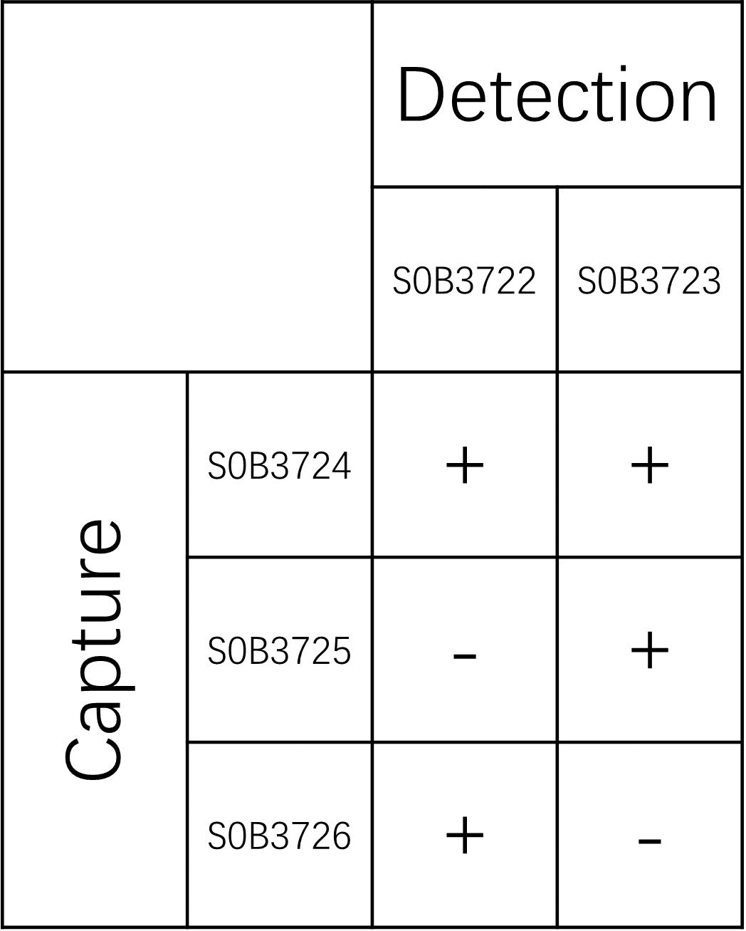
| Application | Sandwich ELISA |
| Purification | Protein A |
| Concentration | 2 mg/ml |
| Conjugation | Unconjugated |
| Physical Appearance | Liquid |
| Storage Buffer | PBS pH7.4, 0.03% Proclin 300 |
| Stability & Storage | 12 months from date of receipt, 2 to 8 °C as supplied |
Background
SLP-76 (SH2 domain-containing leukocyte protein of 76 kDa), specifically when phosphorylated at serine 376 (pS376), is a critical adaptor protein in T-cell receptor (TCR) signaling pathways that plays a pivotal role in immune cell activation and downstream signal transduction. Located within the C-terminal region of the SLP-76 molecule, the phosphorylation of serine 376 serves as a key regulatory switch that modulates the protein’s stability, subcellular localization, and interaction with other signaling molecules such as Vav1 and Itk, thereby influencing actin cytoskeleton reorganization and immunological synapse formation. This specific post-translational modification is often induced upon TCR engagement and co-stimulation, facilitating the assembly of multi-protein signaling complexes that amplify calcium flux and MAPK pathway activation, ultimately leading to T-cell proliferation, cytokine production, and differentiation; consequently, the detection and analysis of SLP-76 pS376 are frequently utilized in immunological research to assess the intensity and quality of T-cell activation states, making it a significant biomarker for understanding both normal immune responses and pathological conditions such as autoimmunity or immunodeficiency.
Picture
Picture
Paired Recommendations