WB result of SLC43A2 Recombinant Rabbit mAb
Primary antibody: SLC43A2 Recombinant Rabbit mAb at 1/1000 dilution
Lane 1: unboiled mouse kidney lysate 20 μg
Lane 2: unboiled mouse liver lysate 20 μg
Secondary antibody: Goat Anti-Rabbit IgG (H+L), HRP conjugated at 1/10000 dilution
Predicted MW: 63 kDa
Observed MW: 63 kDa
SLC43A2 Recombinant Rabbit mAb (S-4492-33)
SLC43A2 Recombinant Rabbit mAb (S-4492-33)
Price:
Regular price
$100 USD
Regular price
Sale price
$100 USD
Unit price
per
For shipping services or bulk orders, you may request a quotation.
Secure checkout with

View full details
Product Details
Product Details
Product Specification
| Host | Rabbit |
| Antigen | SLC43A2 |
| Immunogen | Synthetic Peptide |
| Location | Cell membrane |
| Accession | Q8N370 |
| Clone Number | S-4492-33 |
| Antibody Type | Recombinant mAb |
| Isotype | IgG |
| Application | WB, IHC-P, IF |
| Reactivity | Hu, Ms |
| Positive Sample | Mouse kidney, mouse liver |
| Predicted Reactivity | Or |
| Purification | Protein A |
| Concentration | 0.5 mg/ml |
| Conjugation | Unconjugated |
| Physical Appearance | Liquid |
| Storage Buffer | PBS, 40% Glycerol, 0.05% BSA, 0.02% sodium azide |
| Stability & Storage | 12 months from date of receipt / reconstitution, -20 °C as supplied |
Dilution
| application | dilution | species |
| WB | 1:1000 | Hu, Ms |
| IHC-P | 1:1000-1:2000 | Hu, Ms |
| IF | 1:500 | Ms |
Background
SLC43A2 (solute carrier family 43 member 2, also known as LAT4) is a sodium-, chloride-, and pH-independent L-type amino acid transporter encoded by the SLC43A2 gene located on human chromosome 17p13.3, containing 569 amino acids with a molecular weight of about 62.7 kDa. It has 12 transmembrane domains and exists as a homodimer on the cell membrane. This protein primarily mediates facilitated diffusion of large neutral amino acids (such as leucine, phenylalanine, methionine, and valine), is highly expressed in the placenta, kidney, and small intestine, and is responsible for transepithelial amino acid transport and reabsorption. Its transport activity is regulated by the phosphorylation status of Ser-274 and Ser-297. Recent studies have revealed a key role for SLC43A2 in tumor immunity: tumor cells overexpressing SLC43A2 competitively take up methionine from CD8⁺ T cells, leading to decreased intracellular methionine and methyl donor SAM levels in T cells, which in turn causes loss of histone H3K79me2, reduced STAT5 expression, and T cell exhaustion. Genetic or biochemical inhibition of tumor SLC43A2 can restore T cell H3K79me2 levels and enhance antitumor immune responses, making SLC43A2 a promising target for cancer immunotherapy. In hepatocellular carcinoma, high SLC43A2 expression is positively correlated with increased infiltration of Tregs, macrophages, and MDSCs and upregulated expression of immune checkpoint molecules, and predicts poor prognosis.
Picture
Picture
Western Blot
Immunohistochemistry
IHC shows positive staining in paraffin-embedded human kidney. Anti-SLC43A2 antibody was used at 1/2000 dilution, followed by a HRP Polymer for Rabbit IgG (ready to use). Counterstained with hematoxylin. Heat mediated antigen retrieval with Tris/EDTA buffer pH9.0 was performed before commencing with IHC staining protocol.
IHC shows positive staining in paraffin-embedded human placenta. Anti-SLC43A2 antibody was used at 1/2000 dilution, followed by a HRP Polymer for Rabbit IgG (ready to use). Counterstained with hematoxylin. Heat mediated antigen retrieval with Tris/EDTA buffer pH9.0 was performed before commencing with IHC staining protocol.
IHC shows positive staining in paraffin-embedded human lung cancer. Anti-SLC43A2 antibody was used at 1/2000 dilution, followed by a HRP Polymer for Rabbit IgG (ready to use). Counterstained with hematoxylin. Heat mediated antigen retrieval with Tris/EDTA buffer pH9.0 was performed before commencing with IHC staining protocol.
IHC shows positive staining in paraffin-embedded human esophageal cancer. Anti-SLC43A2 antibody was used at 1/1000 dilution, followed by a HRP Polymer for Rabbit IgG (ready to use). Counterstained with hematoxylin. Heat mediated antigen retrieval with Tris/EDTA buffer pH9.0 was performed before commencing with IHC staining protocol.
Negative control: IHC shows negative staining in paraffin-embedded human cardiac muscle. Anti-SLC43A2 antibody was used at 1/2000 dilution, followed by a HRP Polymer for Rabbit IgG (ready to use). Counterstained with hematoxylin. Heat mediated antigen retrieval with Tris/EDTA buffer pH9.0 was performed before commencing with IHC staining protocol.
IHC shows positive staining in paraffin-embedded mouse liver. Anti-SLC43A2 antibody was used at 1/2000 dilution, followed by a HRP Polymer for Rabbit IgG (ready to use). Counterstained with hematoxylin. Heat mediated antigen retrieval with Tris/EDTA buffer pH9.0 was performed before commencing with IHC staining protocol.
Immunofluorescence
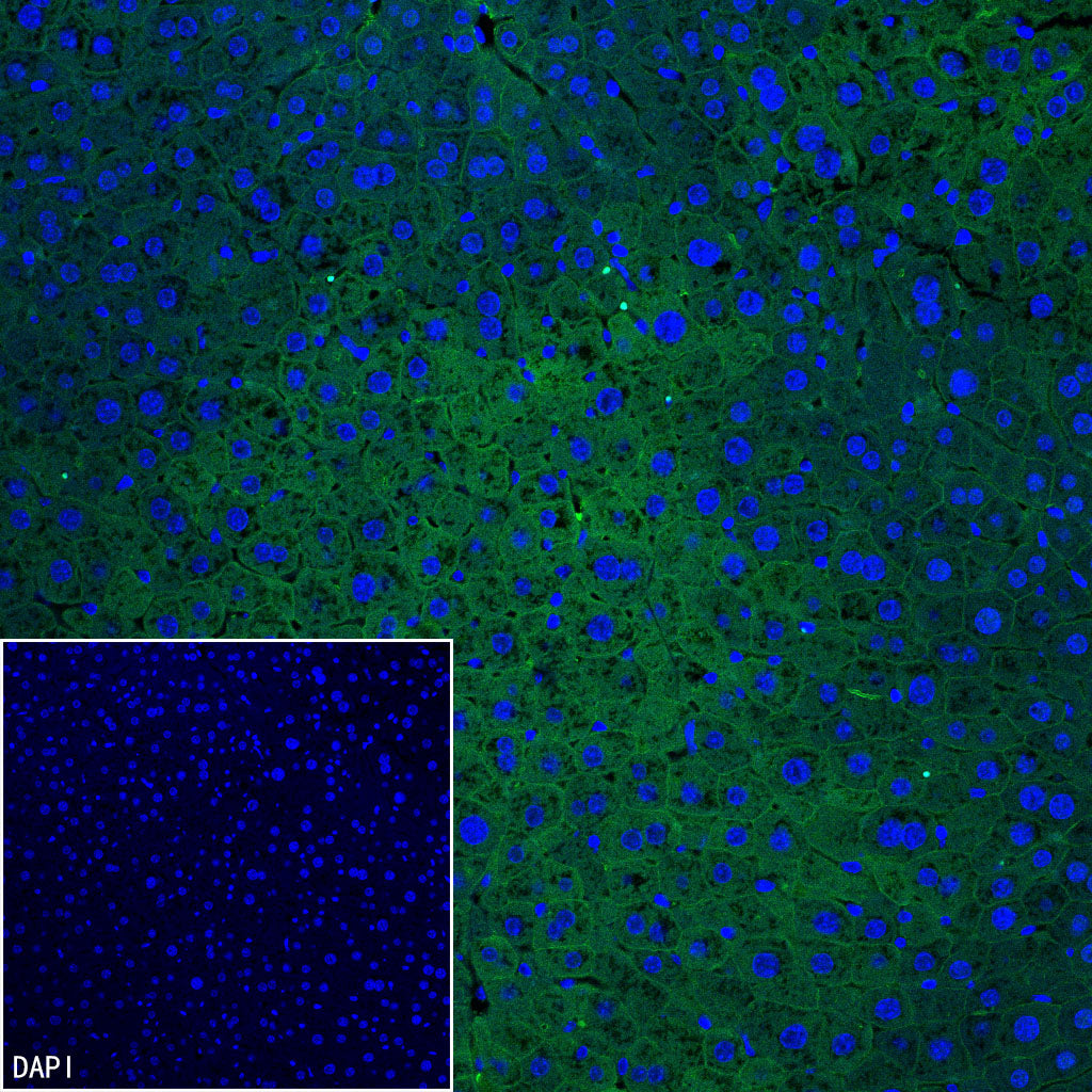
IF shows positive staining in paraffin-embedded mouse liver. Anti-SLC43A2 antibody was used at 1/500 dilution (Green) and incubated overnight at 4°C. Goat polyclonal Antibody to Rabbit IgG - H&L (Alexa Fluor® 488) was used as secondary antibody at 1/1000 dilution. Counterstained with DAPI (Blue). Heat mediated antigen retrieval with EDTA buffer pH9.0 was performed before commencing with IF staining protocol.