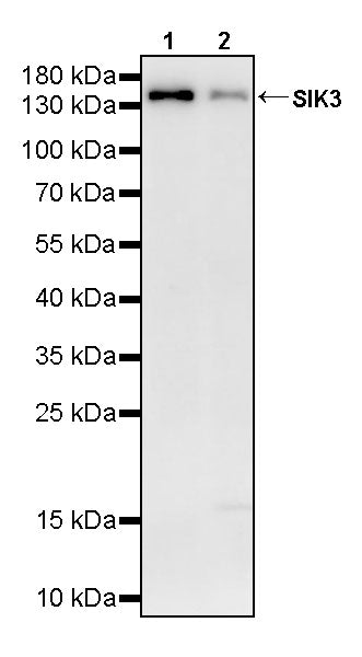
WB result of SIK3 Rabbit pAb
Primary antibody: SIK3 Rabbit pAb at 1/1000 dilution
Lane 1: A-673 whole cell lysate 20 µg
Lane 2: KATO III whole cell lysate 20 µg
Lane 2: U-87 MG whole cell lysate 20 µg
Secondary antibody: Goat Anti-rabbit IgG (H+L), HRP conjugated at 1/10000 dilution
Predicted MW: 145 kDa
Observed MW: 145 kDa
SIK3 Rabbit Polyclonal Antibody
SIK3 Rabbit Polyclonal Antibody
Price:
Regular price
$100 USD
Regular price
Sale price
$100 USD
Unit price
per
For shipping services or bulk orders, you may request a quotation.
Secure checkout with

View full details
Product Details
Product Details
Product Specification
| Host | Rabbit |
| Antigen | SIK3 |
| Synonyms | Serine/threonine-protein kinase SIK3; Salt-inducible kinase 3 (SIK-3); Serine/threonine-protein kinase QSK; KIAA0999; QSK |
| Immunogen | Synthetic Peptide |
| Location | Cytoplasm |
| Accession | Q9Y2K2 |
| Antibody Type | Polyclonal antibody |
| Isotype | IgG |
| Application | WB |
| Reactivity | Hu, Ms, Rt |
| Predicted Reactivity | / |
| Purification | Immunogen Affinity |
| Concentration | 0.5 mg/ml |
| Conjugation | Unconjugated |
| Physical Appearance | Liquid |
| Storage Buffer | PBS, 40% Glycerol, 0.05% BSA, 0.02% sodium azide |
| Stability & Storage | 12 months from date of receipt / reconstitution, -20 °C as supplied |
Dilution
| application | dilution | species |
| WB | 1:1000 | Hu, Ms, Rt |
Background
SIK3 (salt-inducible kinase 3, also known as QSK) is a serine/threonine protein kinase encoded by the SIK3 gene located on chromosome 11q23.3, belonging to the AMPK-related kinase family, and is most abundantly expressed in the brain and testis. The protein has a typical AMPK family domain organization: an N-terminal kinase catalytic domain, a ubiquitin-associated (UBA) domain, and a long C-terminal tail lacking obvious structural features. The catalytic activity of SIK3 depends on constitutive phosphorylation of Thr221 in its T-loop by the upstream kinase LKB1, whereas protein kinase A (PKA) inhibits its activity by phosphorylating multiple sites in its C-terminus (such as Thr469 and Ser551) and promotes 14-3-3 protein binding to sequester it in the cytoplasm. Functionally, SIK3 first attracted attention for its regulation of sleep—its "Sleepy" mutation causes a significant increase in sleep need in mice, and it participates in the phosphorylation-mediated regulation of sleep homeostasis. At the same time, SIK3 is indispensable for skeletal development: it phosphorylates HDAC4 to sequester it in the cytoplasm, thereby relieving inhibition of Mef2c to promote chondrocyte hypertrophy; loss of its function leads to dwarfism and skeletal malformations, while human SIK3 mutations cause Krakow-type spondyloepimetaphyseal dysplasia. Recent studies have also revealed a key role for SIK3 in Alzheimer's disease: SIK3 influences hippocampal synaptic plasticity and cognitive function by forming a complex with HDAC4 and regulating its nucleocytoplasmic shuttling, and restoring SIK3 activity in AD mouse models improves synaptic function and cognitive deficits. In addition, SIK3 participates in the regulation of cholesterol and bile acid metabolism, circadian rhythms, inflammatory responses, and tumorigenesis, among various other pathophysiological processes.
Picture
Picture
Western Blot
WB result of SIK3 Rabbit pAb
Primary antibody: SIK3 Rabbit pAb at 1/1000 dilution
Lane 1: Neuro-2a whole cell lysate 20 µg
Lane 2: mouse brain lysate 20 µg
Secondary antibody: Goat Anti-rabbit IgG (H+L), HRP conjugated at 1/10000 dilution
Predicted MW: 145 kDa
Observed MW: 145 kDa
WB result of SIK3 Rabbit pAb
Primary antibody: SIK3 Rabbit pAb at 1/1000 dilution
Lane 1: C6 whole cell lysate 20 µg
Lane 2: rat brain lysate 20 µg
Secondary antibody: Goat Anti-rabbit IgG (H+L), HRP conjugated at 1/10000 dilution
Predicted MW: 145 kDa
Observed MW: 145 kDa