WB result of SHIP1 Recombinant Rabbit mAb
Primary antibody: SHIP1 Recombinant Rabbit mAb at 1/1000 dilution
Lane 1: Jurkat whole cell lysate 20 µg
Lane 2: Raji whole cell lysate 20 µg
Lane 3: Daudi whole cell lysate 20 µg
Lane 4: Ramos whole cell lysate 20 µg
Negative control: Jurkat whole cell lysate
Secondary antibody: Goat Anti-rabbit IgG (H+L), HRP conjugated at 1/10000 dilution
Predicted MW: 133 kDa
Observed MW: 133 kDa
This blot was developed with high sensitivity substrate
SHIP1 Recombinant Rabbit mAb (S-4235-32)
SHIP1 Recombinant Rabbit mAb (S-4235-32)
Price:
Regular price
$100 USD
Regular price
Sale price
$100 USD
Unit price
per
For shipping services or bulk orders, you may request a quotation.
Secure checkout with

View full details
Product Details
Product Details
Product Specification
| Host | Rabbit |
| Antigen | SHIP1 |
| Synonyms | Phosphatidylinositol 3,4,5-trisphosphate 5-phosphatase 1; Inositol polyphosphate-5-phosphatase D; Inositol polyphosphate-5-phosphatase of 145 kDa (SIP-145); Phosphatidylinositol 4,5-bisphosphate 5-phosphatase; SH2 domain-containing inositol 5'-phosphatase 1 (SH2 domain-containing inositol phosphatase 1; SHIP-1); p150Ship (hp51CN); SHIP; INPP5D |
| Immunogen | Synthetic Peptide |
| Location | Cell membrane |
| Accession | Q92835 |
| Clone Number | S-4235-32 |
| Antibody Type | Recombinant mAb |
| Isotype | IgG |
| Application | WB, IHC-P |
| Reactivity | Hu |
| Purification | Protein A |
| Concentration | 2 mg/ml |
| Conjugation | Unconjugated |
| Physical Appearance | Liquid |
| Storage Buffer | PBS, 40% Glycerol, 0.05% BSA, 0.02% sodium azide |
| Stability & Storage | 12 months from date of receipt / reconstitution, -20 °C as supplied |
Dilution
| application | dilution | species |
| WB | 1:1000 | Hu |
| IHC-P | 1:800 | Hu |
Background
SHIP1 (SH2 domain-containing inositol polyphosphate 5-phosphatase 1, encoded by the INPP5D gene) is a lipid phosphatase primarily expressed in hematopoietic cells that serves as a negative regulator of the PI3K signaling pathway, catalyzing the dephosphorylation of PI(3,4,5)P3 to PI(3,4)P2 and thereby terminating activation signals downstream of effectors such as Akt. The protein is a multi-domain protein of approximately 145 kDa, comprising an N-terminal SH2 domain, a PH-related domain, a central 5-phosphatase catalytic domain, a C2 domain, and an intrinsically disordered C-terminal domain rich in NPxY and proline motifs. SHIP1 has an autoinhibitory mechanism: the N-terminal SH2 domain forms intramolecular contacts with the CBL1 motif of the C2 domain, sterically hindering membrane binding by the C2 domain and thereby limiting its phosphatase activity; when the SH2 domain binds to phosphotyrosine peptides derived from immunoreceptor ITIM motifs, autoinhibition is relieved, allowing SHIP1 to translocate to the plasma membrane and exert its catalytic function. Functionally, SHIP1 is a key mediator of inhibitory signaling in B cells, and its loss causes a chronic myeloid leukemia-like myeloproliferative disease in mice. In recent years, the role of SHIP1 in the central nervous system has attracted increasing attention: it is highly expressed in microglia and regulates complement-mediated synaptic pruning, and its loss leads to excessive synaptic engulfment early in development and cognitive deficits in adulthood. In addition, INPP5D is a risk gene for Alzheimer's disease, and abnormal SHIP1 function is associated with microglial inflammatory dysregulation and neurodegeneration. SHIP1 also participates in platelet activation and thrombosis, regulation of allergic responses, and the pathological processes of inflammatory diseases such as Crohn's disease. Given its broad functions, SHIP1 inhibitors (such as 3AC) and activators (such as AQX-435) are being developed for therapeutic exploration in leukemia, Alzheimer's disease, and inflammatory diseases.
Picture
Picture
Western Blot
Immunohistochemistry
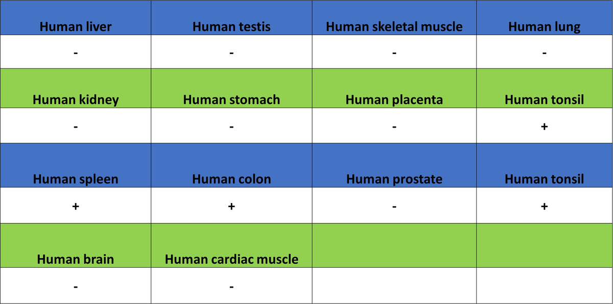
IHC shows positive staining in paraffin-embedded human spleen. Anti-SHIP1 antibody was used at 1/800 dilution, followed by a HRP Polymer for Rabbit IgG (ready to use). Counterstained with hematoxylin. Heat mediated antigen retrieval with Tris/EDTA buffer pH9.0 was performed before commencing with IHC staining protocol.
IHC shows positive staining in paraffin-embedded human tonsil. Anti-SHIP1 antibody was used at 1/800 dilution, followed by a HRP Polymer for Rabbit IgG (ready to use). Counterstained with hematoxylin. Heat mediated antigen retrieval with Tris/EDTA buffer pH9.0 was performed before commencing with IHC staining protocol.
Negative control: IHC shows negative staining in paraffin-embedded human liver. Anti-SHIP1 antibody was used at 1/800 dilution, followed by a HRP Polymer for Rabbit IgG (ready to use). Counterstained with hematoxylin. Heat mediated antigen retrieval with Tris/EDTA buffer pH9.0 was performed before commencing with IHC staining protocol.