WB result of SF3B1 Rabbit Polyclonal Antibody
Primary antibody: SF3B1 Rabbit Polyclonal Antibody at 1/1000 dilution
Lane 1: HeLa whole cell lysate 20 μg
Lane 2: Jurkat whole cell lysate 20 μg
Lane 3: K-562 whole cell lysate 20 μg
Lane 4: Ramos whole cell lysate 20 μg
Lane 5: PANC-1 whole cell lysate 20 μg
Secondary antibody: Goat Anti-Rabbit IgG (H+L), HRP conjugated at 1/10000 dilution
Predicted MW: 146 kDa
Observed MW: 155 kDa
SF3B1 Rabbit Polyclonal Antibody
SF3B1 Rabbit Polyclonal Antibody
Price:
Regular price
$100 USD
Regular price
Sale price
$100 USD
Unit price
per
For shipping services or bulk orders, you may request a quotation.
Secure checkout with

View full details
Product Details
Product Details
Product Specification
| Host | Rabbit |
| Antigen | SF3B1 |
| Immunogen | Synthetic Peptide |
| Location | Cytoplasm, Nucleus |
| Accession | O75533 |
| Antibody Type | Polyclonal antibody |
| Isotype | IgG |
| Application | WB, IHC-P, ICC |
| Reactivity | Hu, Ms, Rt |
| Positive Sample | HeLa, Jurkat, K-562, Ramos, PANC-1, NIH/3T3, RAW264.7, mouse brain, PC-12, C6, rat brain |
| Purification | Immunogen Affinity |
| Concentration | 0.5 mg/ml |
| Conjugation | Unconjugated |
| Physical Appearance | Liquid |
| Storage Buffer | PBS, 40% Glycerol, 0.05% BSA, 0.02% sodium azide |
| Stability & Storage | 12 months from date of receipt / reconstitution, -20 °C as supplied |
Dilution
| application | dilution | species |
| WB | 1:1000-1:5000 | Hu, Ms, Rt |
| IHC-P | 1:500 | Hu, Ms, Rt |
| ICC | 1:500 | Hu, Ms, Rt |
Background
SF3B1 (splicing factor 3B subunit 1) is a core component of the U2 small nuclear ribonucleoprotein complex, encoded by the SF3B1 gene located on human chromosome 2q33.1, containing approximately 1,304 amino acids with a molecular weight of about 145.8 kDa. Its structure comprises multiple coiled-coil regions, disordered regions, and a C-terminal HEAT repeat domain, and it interacts with the RNA recognition motif of p14 protein through a C-terminal β-strand; it is essential for the assembly of the "A" spliceosomal complex (in which U2 snRNP stably binds the pre-mRNA branch point sequence) and the "E" spliceosomal complex. Functionally, SF3B1 is responsible for recognizing the branch point adenosine during pre-mRNA splicing and coordinating spliceosome assembly. Recent cryo-electron microscopy studies have further revealed that the SF3B complex binds its targets by recognizing specific RNA shapes rather than sequences, a mechanism that applies to both pre-mRNA splicing and nuclear export of mature mRNA. Mutations in SF3B1 are high-frequency driver events in multiple tumors, with K700E and R625H being hotspot mutations; these mutations remodel the splicing factor network through aberrant recruitment of factors such as SUGP1 and DHX15, activate cryptic splice sites, and thereby affect genes related to RNA metabolism, cell cycle, and genomic stability, as well as triggering downstream signaling pathways such as NF-κB, AKT, and p53 to promote tumorigenesis. In myelodysplastic syndromes, SF3B1 mutations are found in approximately 28% of patients, are highly correlated with the ring sideroblast phenotype (positive predictive value 97.7%), and independently predict better overall survival and a lower risk of transformation to acute myeloid leukemia. In addition, SF3B1 mutations are also found in chronic lymphocytic leukemia, breast cancer, and uveal melanoma, and spliceosome inhibitors such as pladienolide B are being explored to target this pathway.
Picture
Picture
Western Blot
WB result of SF3B1 Rabbit Polyclonal Antibody
Primary antibody: SF3B1 Rabbit Polyclonal Antibody at 1/1000 dilution
Lane 1: NIH/3T3 whole cell lysate 20 μg
Lane 2: RAW 264.7 whole cell lysate 20 μg
Lane 3: mouse brain lysate 20 μg
Secondary antibody: Goat Anti-Rabbit IgG (H+L), HRP conjugated at 1/10000 dilution
Predicted MW: 146 kDa
Observed MW: 155 kDa
WB result of SF3B1 Rabbit Polyclonal Antibody
Primary antibody: SF3B1 Rabbit Polyclonal Antibody at 1/1000 dilution
Lane 1: PC-12 whole cell lysate 20 μg
Lane 2: C6 whole cell lysate 20 μg
Lane 3: rat brain lysate 20 μg
Secondary antibody: Goat Anti-Rabbit IgG (H+L), HRP conjugated at 1/10000 dilution
Predicted MW: 146 kDa
Observed MW: 155 kDa
Immunohistochemistry
IHC shows positive staining in paraffin-embedded human brain. Anti-SF3B1 antibody was used at 1/500 dilution, followed by a HRP Polymer for Rabbit IgG (ready to use). Counterstained with hematoxylin. Heat mediated antigen retrieval with Tris/EDTA buffer pH9.0 was performed before commencing with IHC staining protocol.
IHC shows positive staining in paraffin-embedded human colon. Anti-SF3B1 antibody was used at 1/500 dilution, followed by a HRP Polymer for Rabbit IgG (ready to use). Counterstained with hematoxylin. Heat mediated antigen retrieval with Tris/EDTA buffer pH9.0 was performed before commencing with IHC staining protocol.
IHC shows positive staining in paraffin-embedded human cervical squamous cell carcinoma. Anti-SF3B1 antibody was used at 1/500 dilution, followed by a HRP Polymer for Rabbit IgG (ready to use). Counterstained with hematoxylin. Heat mediated antigen retrieval with Tris/EDTA buffer pH9.0 was performed before commencing with IHC staining protocol.
IHC shows positive staining in paraffin-embedded human lung cancer. Anti-SF3B1 antibody was used at 1/500 dilution, followed by a HRP Polymer for Rabbit IgG (ready to use). Counterstained with hematoxylin. Heat mediated antigen retrieval with Tris/EDTA buffer pH9.0 was performed before commencing with IHC staining protocol.
IHC shows positive staining in paraffin-embedded mouse spleen. Anti-SF3B1 antibody was used at 1/500 dilution, followed by a HRP Polymer for Rabbit IgG (ready to use). Counterstained with hematoxylin. Heat mediated antigen retrieval with Tris/EDTA buffer pH9.0 was performed before commencing with IHC staining protocol.
IHC shows positive staining in paraffin-embedded rat testis. Anti-SF3B1 antibody was used at 1/500 dilution, followed by a HRP Polymer for Rabbit IgG (ready to use). Counterstained with hematoxylin. Heat mediated antigen retrieval with Tris/EDTA buffer pH9.0 was performed before commencing with IHC staining protocol.
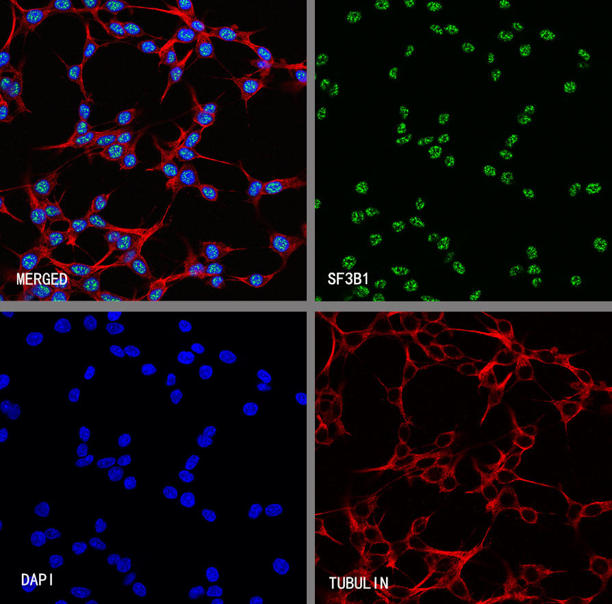
Immunocytochemistry
ICC shows positive staining in HeLa cells. Anti- SF3B1 antibody was used at 1/500 dilution (Green) and incubated overnight at 4°C. Goat polyclonal Antibody to Rabbit IgG - H&L (Alexa Fluor® 488) was used as secondary antibody at 1/1000 dilution. The cells were fixed with 4% PFA and permeabilized with 0.1% PBS-Triton X-100. Nuclei were counterstained with DAPI (Blue). Counterstain with tubulin (Red).
ICC shows positive staining in RAW 264.7 cells. Anti- SF3B1 antibody was used at 1/500 dilution (Green) and incubated overnight at 4°C. Goat polyclonal Antibody to Rabbit IgG - H&L (Alexa Fluor® 488) was used as secondary antibody at 1/1000 dilution. The cells were fixed with 4% PFA and permeabilized with 0.1% PBS-Triton X-100. Nuclei were counterstained with DAPI (Blue). Counterstain with tubulin (Red).
ICC shows positive staining in C6 cells. Anti- SF3B1 antibody was used at 1/500 dilution (Green) and incubated overnight at 4°C. Goat polyclonal Antibody to Rabbit IgG - H&L (Alexa Fluor® 488) was used as secondary antibody at 1/1000 dilution. The cells were fixed with 100% ice-cold methanol and permeabilized with 0.1% PBS-Triton X-100. Nuclei were counterstained with DAPI (Blue). Counterstain with tubulin (Red).