Serum Amyloid A Recombinant Rabbit mAb (S-5308)
Serum Amyloid A Recombinant Rabbit mAb (S-5308)
Price:
Regular price
$100 USD
Regular price
Sale price
$100 USD
Unit price
per
For shipping services or bulk orders, you may request a quotation.
Secure checkout with

View full details
Product Details
Product Details
Product Specification
| Host | Rabbit |
| Antigen | Serum Amyloid A |
| Synonyms | Serum amyloid A-1 protein; Amyloid fibril protein AA; Serum amyloid protein A(2-104); Serum amyloid protein A(3-104); Serum amyloid protein A(2-103); Serum amyloid protein A(2-102); Serum amyloid protein A(4-101); SAA1 |
| Location | Secreted |
| Accession | P0DJI8 |
| Clone Number | S-5308 |
| Antibody Type | Recombinant mAb |
| Isotype | IgG |
| Application | WB, IHC-P |
| Reactivity | Hu |
| Positive Sample | Human plasma, Human serum |
| Purification | Protein A |
| Concentration | 0.5 mg/ml |
| Conjugation | Unconjugated |
| Physical Appearance | Liquid |
| Storage Buffer | PBS, 40% Glycerol, 0.05% BSA, 0.02% sodium azide |
| Stability & Storage | 12 months from date of receipt / reconstitution, -20 °C as supplied |
Dilution
| application | dilution | species |
| WB | 1:500-1:1000 | Hu |
| IHC-P | 1:1000 | Hu |
Background
Serum Amyloid A (SAA) is an acute-phase reactant protein synthesized by hepatocytes and secreted into the bloodstream. Under normal physiological conditions, its serum concentration is extremely low; however, during acute-phase responses such as infection, inflammation, or tissue injury, its levels can surge dramatically by hundreds to thousands of times within hours. Structurally, SAA is an apolipoprotein that primarily associates with high-density lipoprotein (HDL) and can interact with cholesterol and phospholipids through its N-terminal α-helical domain, thereby participating in the regulation of lipid metabolism. Functionally, SAA plays multifaceted roles in immune defense and inflammatory regulation: it can recruit immune cells such as monocytes and neutrophils to sites of inflammation through chemotaxis, induce the release of various pro-inflammatory cytokines, and participate in antigen presentation and immune response modulation. However, dysregulation of SAA function is closely associated with various diseases; persistently elevated SAA levels serve as a biomarker for chronic inflammatory diseases such as rheumatoid arthritis and atherosclerosis. In amyloidosis, proteolytic fragments of SAA can deposit in tissues to form amyloid A (AA) fibrils, leading to multi-organ dysfunction. Additionally, within the tumor microenvironment, SAA has been found to promote tumor invasion and metastasis. Due to its high sensitivity to inflammation, SAA has become an important serological marker for the differential diagnosis of infection and inflammation in clinical practice.
Picture
Picture
Western Blot
WB result of Serum Amyloid A Recombinant Rabbit mAb
Primary antibody: Serum Amyloid A Recombinant Rabbit mAb at 1/1000 dilution
Lane 1: Human plasma lysate 20 µg
Lane 2: Human serum lysate 20 µg
Secondary antibody: Goat Anti-rabbit IgG, (H+L), HRP conjugated at 1/10000 dilution
Predicted MW: 12 kDa
Observed MW: 14 kDa
Immunohistochemistry
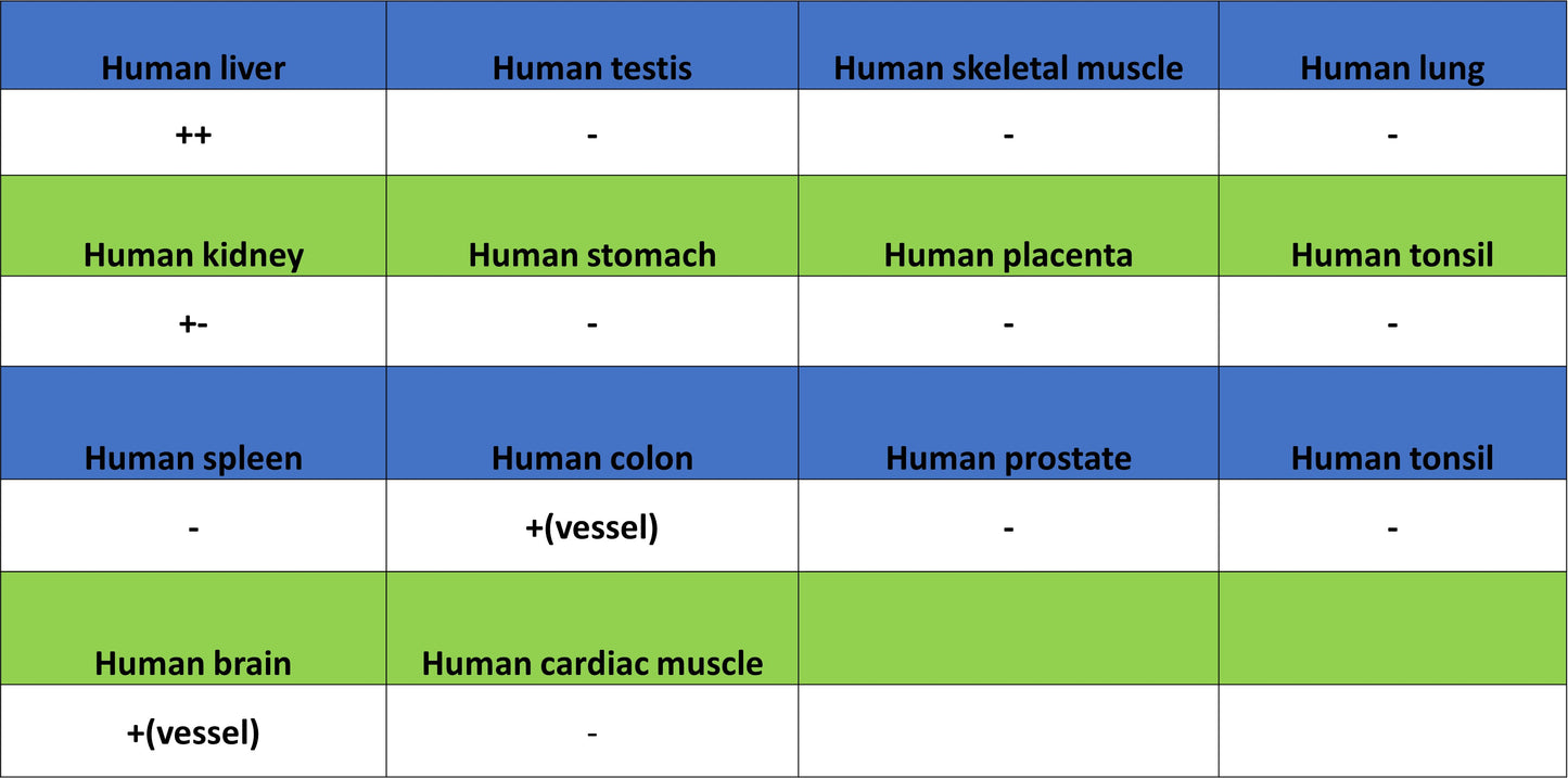
IHC shows positive staining in paraffin-embedded human liver. Anti-Serum Amyloid A antibody was used at 1/1000 dilution, followed by a HRP Polymer for Rabbit IgG (ready to use). Counterstained with hematoxylin. Heat mediated antigen retrieval with Tris/EDTA buffer pH9.0 was performed before commencing with IHC staining protocol.