WB result of SAE2 Recombinant Rabbit mAb
Primary antibody: SAE2 Recombinant Rabbit mAb at 1/1000 dilution
Lane 1: HeLa whole cell lysate 20 μg
Lane 2: A549 whole cell lysate 20 μg
Lane 3: K-562 whole cell lysate 20 μg
Lane 4: HT-29 whole cell lysate 20 μg
Secondary antibody: Goat Anti-Rabbit IgG (H+L), HRP conjugated at 1/10000 dilution
Predicted MW: 71 kDa
Observed MW: 100 kDa
SAE2 Recombinant Rabbit mAb (S-4888-5)
SAE2 Recombinant Rabbit mAb (S-4888-5)
Price:
Regular price
$100 USD
Regular price
Sale price
$100 USD
Unit price
per
For shipping services or bulk orders, you may request a quotation.
Secure checkout with

View full details
Product Details
Product Details
Product Specification
| Host | Rabbit |
| Antigen | SAE2 |
| Immunogen | Synthetic Peptide |
| Location | Cytoplasm, Nucleus |
| Accession | Q9UBT2 |
| Clone Number | S-4888-5 |
| Antibody Type | Recombinant mAb |
| Isotype | IgG |
| Application | WB, ICC |
| Reactivity | Hu, Ms, Rt, Mk |
| Positive Sample | HeLa, A549, K-562, HT-29, Raw 264.7, mouse brain, PC-12, COS-7 |
| Predicted Reactivity | Zf |
| Purification | Protein A |
| Concentration | 0.5 mg/ml |
| Conjugation | Unconjugated |
| Physical Appearance | Liquid |
| Storage Buffer | PBS, 40% Glycerol, 0.05% BSA, 0.02% sodium azide |
| Stability & Storage | 12 months from date of receipt / reconstitution, -20 °C as supplied |
Dilution
| application | dilution | species |
| WB | 1:1000-1:5000 | Hu, Ms, Rt, Mk |
| ICC | 1:500 | Hu, Ms |
Background
SAE2 (also known as UBA2) is the catalytic subunit of the sole E1 activating enzyme in the SUMOylation pathway, forming an obligatory heterodimer with the regulatory subunit SAE1, with a molecular weight of approximately 70 kDa. Its structure consists of multiple functional domains: the N-terminal adenylation domain is responsible for ATP-dependent adenylation of the SUMO C-terminus; the central Cys domain contains the catalytic cysteine Cys173, which forms a high-energy thioester bond with SUMO; the ubiquitin-fold domain provides the E2-binding surface; and the C-terminal unstructured region contains a nuclear localization signal. In the SUMOylation cascade, SAE1:SAE2 first catalyzes adenylation of the C-terminal glycine of SUMO, then forms a thioester bond between Cys173 of SAE2 and SUMO, and ultimately transfers the activated SUMO to the E2 enzyme UBC9. Notably, the Cys domain of SAE2 can undergo autosumoylation, which inhibits its normal interaction with E2 and thereby negatively regulates SUMOylation efficiency; de-SUMOylation of E1 under heat shock conditions is associated with elevated global SUMOylation levels. The nucleocytoplasmic shuttling and nuclear accumulation of SAE2 depend on SUMOylation of multiple lysine residues within its C-terminal nuclear localization signal. A 2025 study further revealed that SAE2 undergoes HDAC6-dependent deacetylation at K164, which in early mitosis promotes conjugation of SUMO1 rather than SUMO2/3, ensuring SUMO1ylation of NuMA to maintain bipolar spindle formation; the acetylation-mimetic mutant SAE2-K164Q preferentially activates SUMO2, leading to multipolar spindles and chromosome segregation defects. In terms of disease associations, UBA2 is highly expressed in various tumors including renal cell carcinoma, glioma, lung cancer, gastric cancer, and colorectal cancer, where it promotes tumor progression, and its variants can also cause a syndrome with congenital skin aplasia and missing digits.
Picture
Picture
Western Blot
WB result of SAE2 Recombinant Rabbit mAb
Primary antibody: SAE2 Recombinant Rabbit mAb at 1/1000 dilution
Lane 1: Raw 264.7 whole cell lysate 20 μg
Lane 2: mouse brain lysate 20 μg
Secondary antibody: Goat Anti-Rabbit IgG (H+L), HRP conjugated at 1/10000 dilution
Predicted MW: 71 kDa
Observed MW: 95 kDa
WB result of SAE2 Recombinant Rabbit mAb
Primary antibody: SAE2 Recombinant Rabbit mAb at 1/1000 dilution
Lane 1: PC-12 whole cell lysate 20 μg
Lane 2: rat brain lysate 20 μg
Secondary antibody: Goat Anti-Rabbit IgG (H+L), HRP conjugated at 1/10000 dilution
Predicted MW: 71 kDa
Observed MW: 95 kDa
WB result of SAE2 Recombinant Rabbit mAb
Primary antibody: SAE2 Recombinant Rabbit mAb at 1/1000 dilution
Lane 1: COS-7 whole cell lysate 20 μg
Secondary antibody: Goat Anti-Rabbit IgG (H+L), HRP conjugated at 1/10000 dilution
Predicted MW: 71 kDa
Observed MW: 95 kDa
Immunocytochemistry
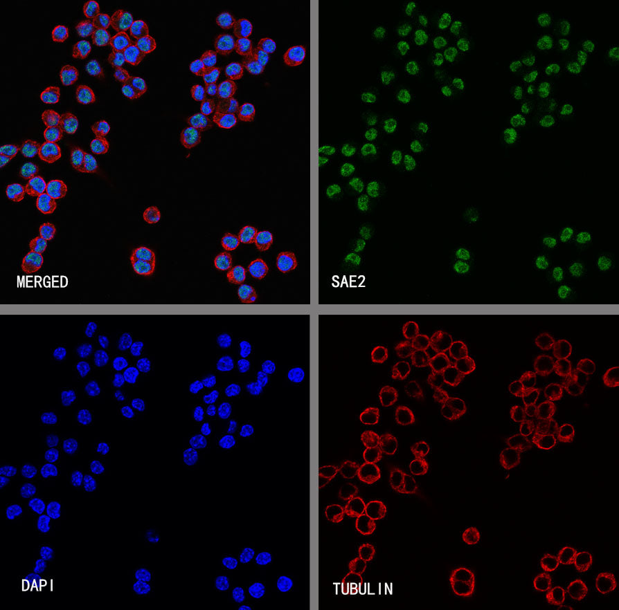
ICC shows positive staining in HeLa cells. Anti- SAE2 antibody was used at 1/500 dilution (Green) and incubated overnight at 4°C. Goat polyclonal Antibody to Rabbit IgG - H&L (Alexa Fluor® 488) was used as secondary antibody at 1/1000 dilution. The cells were fixed with 4% PFA and permeabilized with 0.1% PBS-Triton X-100. Nuclei were counterstained with DAPI (Blue). Counterstain with tubulin (Red).
ICC shows positive staining in RAW 264.7 cells. Anti- SAE2 antibody was used at 1/500 dilution (Green) and incubated overnight at 4°C. Goat polyclonal Antibody to Rabbit IgG - H&L (Alexa Fluor® 488) was used as secondary antibody at 1/1000 dilution. The cells were fixed with 4% PFA and permeabilized with 0.1% PBS-Triton X-100. Nuclei were counterstained with DAPI (Blue). Counterstain with tubulin (Red).