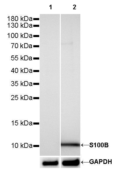
WB result of S100B Rabbit mAb
Primary antibody: S100B Rabbit mAb at 1/5000 dilution
Lane 1: T-47D whole cell lysate 20 µg
Lane 2: SK-MEL-28 whole cell lysate 20 µg
Negative control: T-47D whole cell lysate
Secondary antibody: Goat Anti-Rabbit IgG, (H+L), HRP conjugated at 1/10000 dilution
Predicted MW: 10 kDa
Observed MW: 10 kDa
Product Details
Product Details
Product Specification
| Host | Rabbit |
| Antigen | S100B |
| Synonyms | Protein S100-B, S-100 protein beta chain |
| Immunogen | Recombinant Protein |
| Location | Cytoplasm, Nucleus |
| Accession | P04271 |
| Clone Number | SDT-093-108 |
| Antibody Type | Rabbit mAb |
| Application | WB, IHC-P, IF |
| Reactivity | Hu, Ms, Rt |
| Purification | Protein A |
| Concentration | 0.125 mg/ml |
| Physical Appearance | Liquid |
| Storage Buffer | PBS, 40% Glycerol, 0.05%BSA, 0.03% Proclin 300 |
| Stability & Storage | 12 months from date of receipt / reconstitution, -20 °C as supplied |
Dilution
| application | dilution | species |
| IHC-P | 1:2500 | Hu, Ms, Rt |
| WB | 1:1000-1:5000 | Hu, Ms, Rt |
| IF | 1:1250 | Ms |
Background
S100B is a Ca2+-binding protein, which is secreted from astrocytes. S100B is widely expressed in astrocytes, certain neuronal populations, Schwann cells, myeloid-derived cells, and a few other cell types. S100B is released in the extracellular space in response to glutamate, serotonin, TNF, IL-1β, beta-amyloid peptides, and lysophosphatidic acid. Extracellular S100B exerts both paracrine and autocrine effects on neurons and glia100, through RAGE, and possibly other receptors. The effect of S100B is concentration dependent and can be either trophic or toxic. In humans, high levels of extracellular S100B have been detected under various clinical conditions, for example, brain trauma, ischemia and neurodegenerative, and inflammatory and psychiatric diseases.

Picture
Picture
Western Blot


WB result of S100B Rabbit mAb
Primary antibody: S100B Rabbit mAb at 1/5000 dilution
Lane 1: mouse lung lysate 20 µg
Lane 2: mouse brain lysate 20 µg
Negative control: mouse lung lysate
Secondary antibody: Goat Anti-Rabbit IgG, (H+L), HRP conjugated at 1/10000 dilution
Predicted MW: 10 kDa
Observed MW: 10 kDa

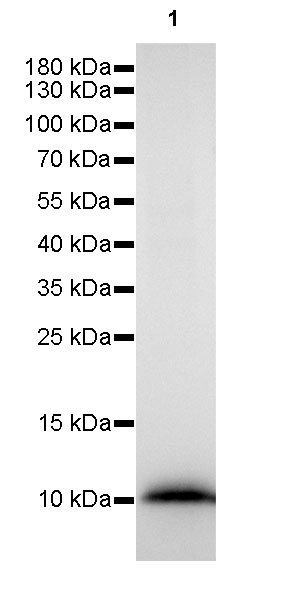
WB result of S100B Rabbit mAb
Primary antibody: S100B Rabbit mAb at 1/5000 dilution
Lane 1: rat brain lysate 20 µg
Secondary antibody: Goat Anti-Rabbit IgG, (H+L), HRP conjugated at 1/10000 dilution
Predicted MW: 10 kDa
Observed MW: 10 kDa
Immunohistochemistry

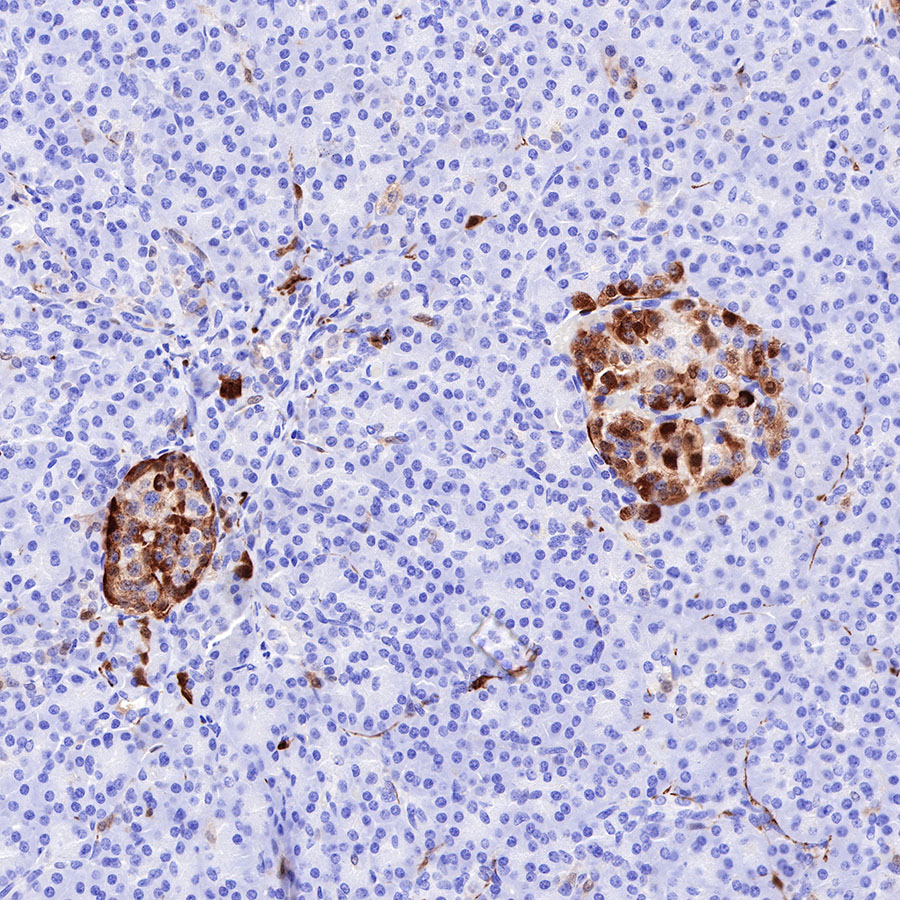
IHC shows positive staining in paraffin-embedded human pancreas. Anti-S100B antibody was used at 1/2500 dilution, followed by a HRP Polymer for Mouse & Rabbit IgG (ready to use). Counterstained with hematoxylin. Heat mediated antigen retrieval with Tris/EDTA buffer pH9.0 was performed before commencing with IHC staining protocol.

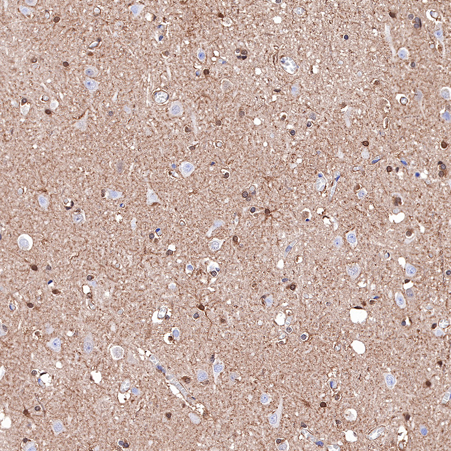
IHC shows positive staining in paraffin-embedded human cerebral cortex. Anti-S100B antibody was used at 1/2500 dilution, followed by a HRP Polymer for Mouse & Rabbit IgG (ready to use). Counterstained with hematoxylin. Heat mediated antigen retrieval with Tris/EDTA buffer pH9.0 was performed before commencing with IHC staining protocol.

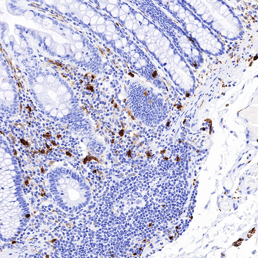
IHC shows positive staining in paraffin-embedded human colon. Anti-S100B antibody was used at 1/2500 dilution, followed by a HRP Polymer for Mouse & Rabbit IgG (ready to use). Counterstained with hematoxylin. Heat mediated antigen retrieval with Tris/EDTA buffer pH9.0 was performed before commencing with IHC staining protocol.

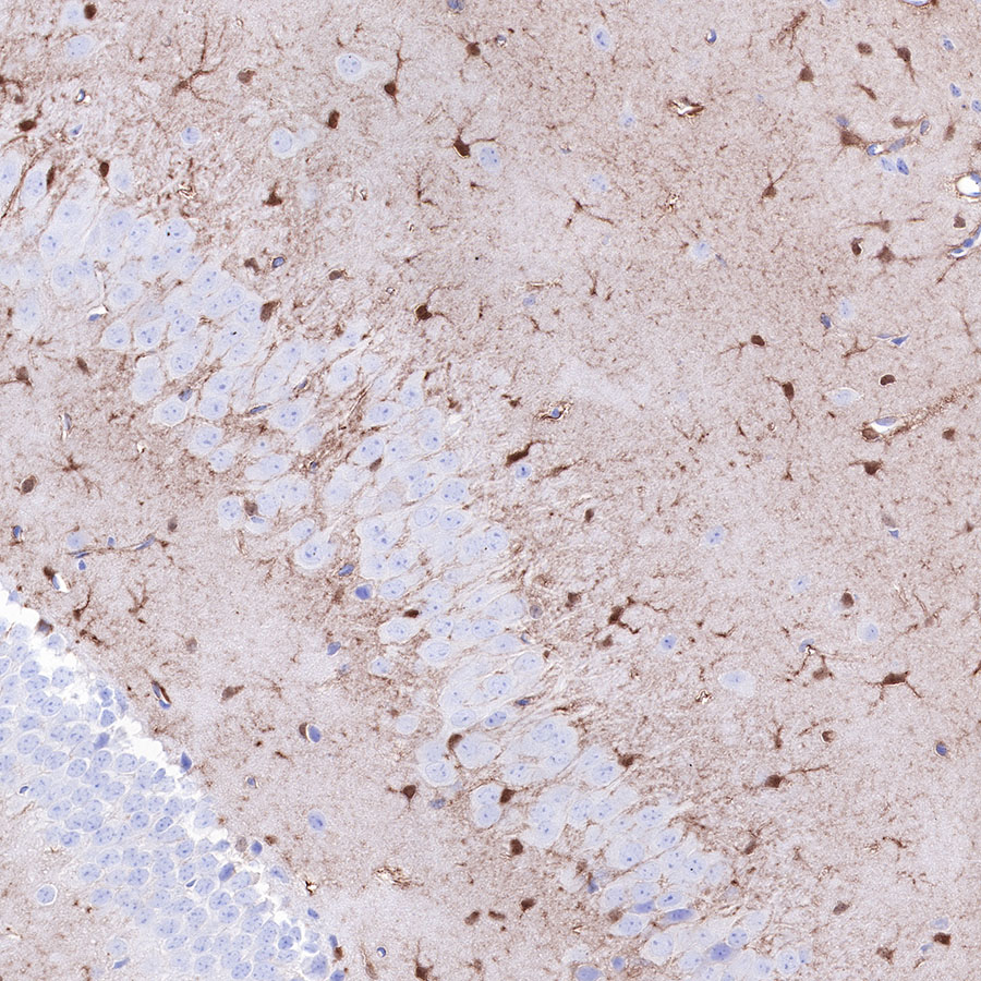
IHC shows positive staining in paraffin-embedded mouse cerebral cortex. Anti-S100B antibody was used at 1/2500 dilution, followed by a HRP Polymer for Mouse & Rabbit IgG (ready to use). Counterstained with hematoxylin. Heat mediated antigen retrieval with Tris/EDTA buffer pH9.0 was performed before commencing with IHC staining protocol.

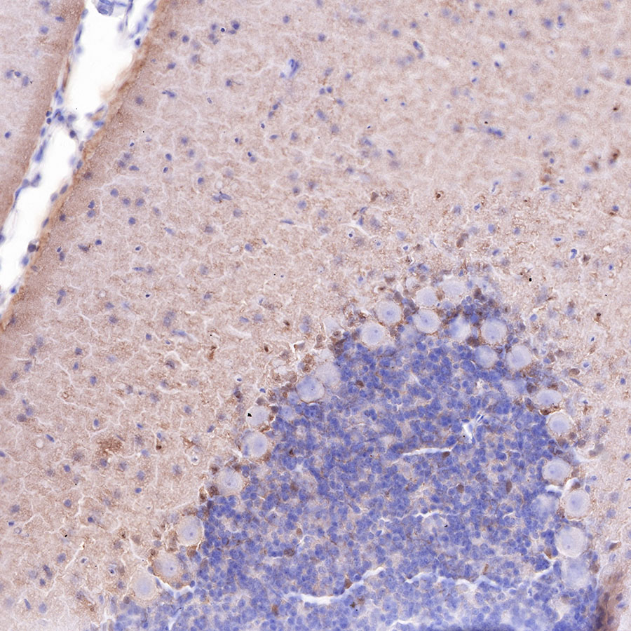
IHC shows positive staining in paraffin-embedded mouse cerebellum. Anti-S100B antibody was used at 1/2500 dilution, followed by a HRP Polymer for Mouse & Rabbit IgG (ready to use). Counterstained with hematoxylin. Heat mediated antigen retrieval with Tris/EDTA buffer pH9.0 was performed before commencing with IHC staining protocol.

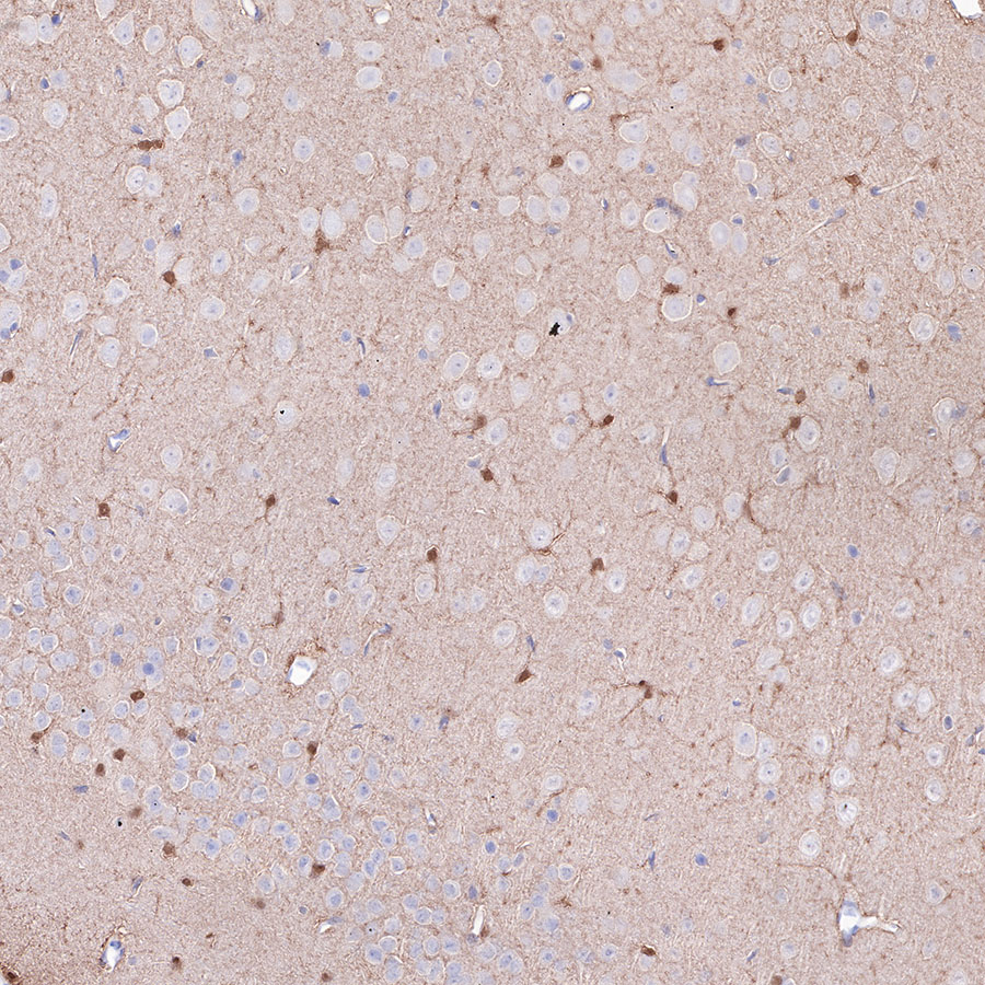
IHC shows positive staining in paraffin-embedded rat cerebral cortex. Anti-S100B antibody was used at 1/2500 dilution, followed by a HRP Polymer for Mouse & Rabbit IgG (ready to use). Counterstained with hematoxylin. Heat mediated antigen retrieval with Tris/EDTA buffer pH9.0 was performed before commencing with IHC staining protocol.

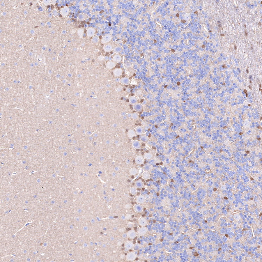
IHC shows positive staining in paraffin-embedded rat cerebellum. Anti-S100B antibody was used at 1/2500 dilution, followed by a HRP Polymer for Mouse & Rabbit IgG (ready to use). Counterstained with hematoxylin. Heat mediated antigen retrieval with Tris/EDTA buffer pH9.0 was performed before commencing with IHC staining protocol.
Immunofluorescence

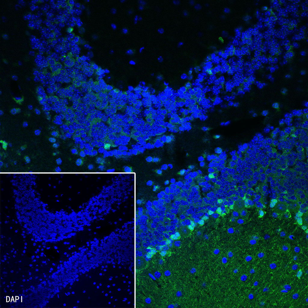
IF shows positive staining in paraffin-embedded mouse cerebellum. Anti-S100B antibody was used at 1/1250 dilution (Green) and incubated overnight at 4°C. Goat polyclonal Antibody to Rabbit IgG - H&L (Alexa Fluor® 488) was used as secondary antibody at 1/1000 dilution. Counterstained with DAPI (Blue). Heat mediated antigen retrieval with Tris/EDTA buffer pH9.0 was performed before commencing with IF staining protocol.