WB result of RXRα Recombinant Rabbit mAb
Primary antibody: RXRα Recombinant Rabbit mAb at 1/2000 dilution
Primary antibody incubation conditions: overnight at 4°C
Lane 1: MCF7 whole cell lysate 20 µg
Lane 2: LNCaP whole cell lysate 20 µg
Lane 3: HeLa whole cell lysate 20 µg
Lane 4: Hep G2whole cell lysate 20 µg
Lane 5: BxPC-3 whole cell lysate 20 µg
Lane 6: HL-60 whole cell lysate 20 µg
Secondary antibody: Goat Anti-rabbit IgG (H+L), HRP conjugated at 1/10000 dilution
Predicted MW: 51 kDa
Observed MW: 55 kDa
RXRα Recombinant Rabbit mAb
RXRα Recombinant Rabbit mAb
Price:
Regular price
$100 USD
Regular price
Sale price
$100 USD
Unit price
per
For shipping services or bulk orders, you may request a quotation.
Secure checkout with

View full details
Product Details
Product Details
Product Specification
| Host | Rabbit |
| Antigen | RXRα |
| Location | Cytoplasm, Nucleus, Mitochondrion |
| Accession | P19793 |
| Antibody Type | Recombinant mAb |
| Isotype | IgG |
| Application | WB, ICC |
| Reactivity | Hu, Ms, Rt |
| Positive Sample | MCF7, LNCaP, HeLa, Hep G2, BxPC-3, HL-60, NIH/3T3, mouse liver, rat liver |
| Purification | Protein A |
| Concentration | 1 mg/ml |
| Conjugation | Unconjugated |
| Physical Appearance | Liquid |
| Storage Buffer | PBS, 40% Glycerol, 0.05% BSA, 0.02% sodium azide |
| Stability & Storage | 12 months from date of receipt / reconstitution, -20 °C as supplied |
Dilution
| application | dilution | species |
| WB | 1:1000-1:2000 | Hu, Ms, Rt |
| ICC | 1:400 | Hu |
Background
RXRα (retinoid X receptor alpha, also known as NR2B1) is a key member of the nuclear receptor superfamily. It consists of approximately 462 amino acids and contains an N-terminal A/B domain, a conserved DNA-binding domain (DBD), a hinge region, and a C-terminal ligand-binding domain (LBD). The DBD mediates recognition of target DNA cis-elements, while the LBD is responsible for ligand binding, dimerization, and AF-2-mediated transcriptional activation. RXRα can form homodimers and homotetramers, and serves as a universal heterodimeric partner for multiple nuclear receptors, including RAR, PPAR, LXR, VDR, TR, and FXR, thereby regulating a broad range of physiological processes such as cell proliferation, differentiation, apoptosis, lipid metabolism, immune inflammation, and embryonic development. Its endogenous ligand is 9-cis retinoic acid, and it can also be modulated by dietary fatty acids such as DHA. Beyond classical nuclear transcriptional regulation, RXRα has important non-genomic functions: it can regulate mitochondrial apoptosis, PI3K/AKT survival signaling, and inflammatory responses in the cytoplasm; in tumor cells, it can be proteolytically cleaved to generate a truncated form, tRXRα, which promotes inflammation and tumor progression; during mitosis, it can be phosphorylated by CDK1 and translocate to the centrosome, where it participates in centrosome assembly through phase separation. Abnormal expression or modification of RXRα is associated with cutaneous T-cell lymphoma, colorectal cancer, metabolic syndrome, and neurodegenerative diseases, and the FDA-approved drug bexarotene (Targretin) is an RXR-based anticancer agent.
Picture
Picture
Western Blot
WB result of RXRα Recombinant Rabbit mAb
Primary antibody: RXRα Recombinant Rabbit mAb at 1/2000 dilution
Primary antibody incubation conditions: overnight at 4°C
Lane 1: NIH/3T3 whole cell lysate 20 µg
Lane 2: mouse liver lysate 20 µg
Secondary antibody: Goat Anti-rabbit IgG (H+L), HRP conjugated at 1/10000 dilution
Predicted MW: 51 kDa
Observed MW: 55 kDa
WB result of RXRα Recombinant Rabbit mAb
Primary antibody: RXRα Recombinant Rabbit mAb at 1/2000 dilution
Primary antibody incubation conditions: overnight at 4°C
Lane 1: rat liver lysate 20 µg
Secondary antibody: Goat Anti-rabbit IgG (H+L), HRP conjugated at 1/10000 dilution
Predicted MW: 51 kDa
Observed MW: 55 kDa
Immunocytochemistry
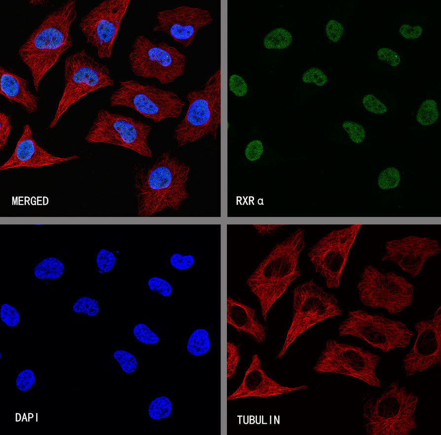
ICC shows positive staining in HeLa cells. Anti-RXRα antibody was used at 1/400 dilution (Green) and incubated overnight at 4°C. Goat polyclonal Antibody to Rabbit IgG - H&L (Alexa Fluor® 488) was used as secondary antibody at 1/1000 dilution. The cells were fixed with 4% PFA and permeabilized with 0.1% PBS-Triton X-100. Nuclei were counterstained with DAPI (Blue). Counterstain with tubulin (Red).