WB result of ROR beta/RORB Recombinant Rabbit mAb
Primary antibody: ROR beta/RORB Recombinant Rabbit mAb at 1/1000 dilution
Lane 1: Y79 whole cell lysate 20 µg
Secondary antibody: Goat Anti- rabbit IgG, (H+L), HRP conjugated at 1/10000 dilution
Predicted MW: 53 kDa
Observed MW: 53 kDa
This blot was developed with high sensitivity substrate.
ROR beta/RORB Recombinant Rabbit mAb (S-5483)
ROR beta/RORB Recombinant Rabbit mAb (S-5483)
Price:
Regular price
$100 USD
Regular price
Sale price
$100 USD
Unit price
per
For shipping services or bulk orders, you may request a quotation.
Secure checkout with

View full details
Product Details
Product Details
Product Specification
| Host | Rabbit |
| Antigen | ROR beta/RORB |
| Synonyms | Nuclear receptor ROR-beta; Nuclear receptor RZR-beta; Nuclear receptor subfamily 1 group F member 2; Retinoid-related orphan receptor-beta; NR1F2; RZRB |
| Location | Nucleus |
| Accession | Q92753 |
| Clone Number | S-5483 |
| Antibody Type | Recombinant mAb |
| Isotype | IgG |
| Application | WB, IHC-P |
| Reactivity | Hu, Ms, Rt |
| Positive Sample | Y79, mouse retina, rat retina |
| Purification | Protein A |
| Concentration | 0.5 mg/ml |
| Conjugation | Unconjugated |
| Physical Appearance | Liquid |
| Storage Buffer | PBS, 40% Glycerol, 0.05% BSA, 0.02% sodium azide |
| Stability & Storage | 12 months from date of receipt / reconstitution, -20 °C as supplied |
Dilution
| application | dilution | species |
| WB | 1:500-1:1000 | Hu, Ms, Rt |
| IHC-P | 1:500 | Ms, Rt |
Background
RORB, also known as RAR-related orphan receptor beta, is a member of the NR1 subfamily within the nuclear receptor superfamily. As a DNA-binding transcription factor, it recognizes and binds to ROR response elements (ROREs) in the promoter regions of downstream genes either as a monomer or homodimer, thereby regulating target gene transcription. Structurally, the protein contains a highly conserved DNA-binding domain (featuring two C4-type zinc fingers) and a C-terminal ligand-binding domain (containing the AF-2 activation function motif), with its transcriptional activity being inhibited by natural ligands such as all-trans retinoic acid (ATRA) through inverse agonism. The functions of RORB are highly tissue-specific and spatiotemporally regulated. In the retina, it is critical for the proper development and differentiation of both rod and cone photoreceptors, regulating photoreceptor cell fate through the induction of transcription factor pathways such as NRL. Additionally, it participates in modulating the periodicity and stability of circadian rhythms, and in the nervous system, it guides cortical neuron architecture and barrel cortex formation. Furthermore, different isoforms of RORB exhibit functional diversity—for example, isoform 1 is indispensable for hindlimb motor control and the differentiation of retinal amacrine and horizontal cells. Loss of function or mutations in the RORB gene have been associated with various diseases, including idiopathic generalized epilepsy (EIG15), myopia, and complex neurodevelopmental disorders.
Picture
Picture
Western Blot
WB result of ROR beta/RORB Recombinant Rabbit mAb
Primary antibody: ROR beta/RORB Recombinant Rabbit mAb at 1/1000 dilution
Lane 1: mouse retina lysate 20 µg
Secondary antibody: Goat Anti- rabbit IgG, (H+L), HRP conjugated at 1/10000 dilution
Predicted MW: 53 kDa
Observed MW: 53 kDa
WB result of ROR beta/RORB Recombinant Rabbit mAb
Primary antibody: ROR beta/RORB Recombinant Rabbit mAb at 1/1000 dilution
Lane 1: rat retina lysate 20 µg
Secondary antibody: Goat Anti- rabbit IgG, (H+L), HRP conjugated at 1/10000 dilution
Predicted MW: 53 kDa
Observed MW: 53 kDa
This blot was developed with high sensitivity substrate.
Immunohistochemistry
IHC shows positive staining in paraffin-embedded mouse retina. Anti-RORB antibody was used at 1/500 dilution, followed by a HRP Polymer for Rabbit IgG (ready to use). Counterstained with hematoxylin. Heat mediated antigen retrieval with Tris/EDTA buffer pH9.0 was performed before commencing with IHC staining protocol.
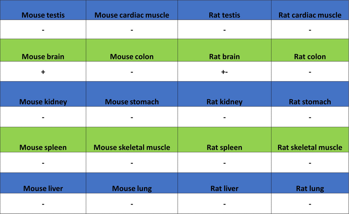
IHC shows positive staining in paraffin-embedded mouse brain. Anti-RORB antibody was used at 1/500 dilution, followed by a HRP Polymer for Rabbit IgG (ready to use). Counterstained with hematoxylin. Heat mediated antigen retrieval with Tris/EDTA buffer pH9.0 was performed before commencing with IHC staining protocol.
IHC shows positive staining in paraffin-embedded rat retina. Anti-RORB antibody was used at 1/500 dilution, followed by a HRP Polymer for Rabbit IgG (ready to use). Counterstained with hematoxylin. Heat mediated antigen retrieval with Tris/EDTA buffer pH9.0 was performed before commencing with IHC staining protocol.
IHC shows positive staining in paraffin-embedded rat brain. Anti-RORB antibody was used at 1/500 dilution, followed by a HRP Polymer for Rabbit IgG (ready to use). Counterstained with hematoxylin. Heat mediated antigen retrieval with Tris/EDTA buffer pH9.0 was performed before commencing with IHC staining protocol.