Ret Recombinant Rabbit mAb (S-4326-45)
Ret Recombinant Rabbit mAb (S-4326-45)
Price:
Regular price
$100 USD
Regular price
Sale price
$100 USD
Unit price
per
For shipping services or bulk orders, you may request a quotation.
Secure checkout with

View full details
Product Details
Product Details
Product Specification
| Host | Rabbit |
| Antigen | Ret |
| Synonyms | Proto-oncogene tyrosine-protein kinase receptor Ret; Cadherin family member 12; Proto-oncogene c-Ret; CDHF12; CDHR16; PTC; RET |
| Immunogen | Recombinant Protein |
| Location | Endosome, Cell membrane |
| Accession | P07949 |
| Clone Number | S-4326-45 |
| Antibody Type | Recombinant mAb |
| Isotype | IgG |
| Application | WB |
| Reactivity | Hu |
| Positive Sample | MCF7, SH-SY5Y, THP-1 |
| Purification | Protein A |
| Concentration | 0.5 mg/ml |
| Conjugation | Unconjugated |
| Physical Appearance | Liquid |
| Storage Buffer | PBS, 40% Glycerol, 0.05% BSA, 0.02% sodium azide |
| Stability & Storage | 12 months from date of receipt / reconstitution, -20 °C as supplied |
Dilution
| application | dilution | species |
| WB | 1:1000 | Hu |
Background
Ret (i.e., rearranged during transfection proto-oncogene-encoded receptor tyrosine kinase, encoded by the RET gene) is a single-pass transmembrane receptor that is critically important in embryonic development and adult tissue homeostasis. Its ligands belong to the glial cell line-derived neurotrophic factor (GDNF) family ligands (GFLs), which require binding to the co-receptor GFRα (GDNF family receptor alpha) before they can engage the extracellular domain of Ret protein, inducing dimerization and autophosphorylation, thereby activating the intracellular tyrosine kinase domain and initiating multiple downstream signaling pathways, including the RAS-MAPK pathway (regulating cell proliferation), the PI3K-AKT pathway (regulating cell survival), and the PLCγ pathway (regulating cell migration and differentiation). Ret protein plays irreplaceable roles in kidney development (promoting ureteric bud branching and nephron formation), enteric nervous system development (guiding neural crest cell migration and differentiation to form intramural enteric plexuses), and the hematopoietic system, with its expression being precisely regulated in a spatiotemporal manner. Gain-of-function mutations in this gene, particularly those involving alterations of extracellular cysteine residues or activating mutations in the intracellular kinase domain, can lead to multiple endocrine neoplasia type 2 (MEN2)—classified as MEN2A (medullary thyroid carcinoma with pheochromocytoma and hyperparathyroidism) and MEN2B (more aggressive medullary thyroid carcinoma with mucosal neuromas and Marfanoid habitus)—as well as familial medullary thyroid carcinoma (FMTC). Conversely, loss-of-function mutations in the RET gene primarily cause Hirschsprung's disease (congenital megacolon), characterized by the absence of ganglion cells in portions of the intestine due to impaired enteric neural crest cell migration, resulting in persistent spastic constriction of the intestinal segment and consequently severe intestinal obstruction. In recent years, selective kinase inhibitors targeting RET (such as Pralsetinib and Selpercatinib) have been successfully translated into clinical practice, providing highly effective targeted therapeutic options for patients with RET-mutant thyroid cancers and non-small cell lung cancers.
Picture
Picture
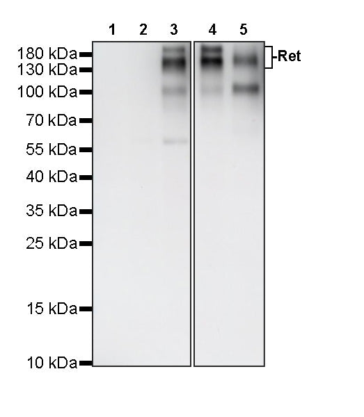
Western Blot
WB result of Ret Recombinant Rabbit mAb
Primary antibody: Ret Recombinant Rabbit mAb at 1/1000 dilution
Lane 1: HepG2 whole cell lysate 20 µg
Lane 2: HeLa whole cell lysate 20 µg
Lane 3: MCF7 whole cell lysate 20 µg
Lane 4: SH-SY5Y whole cell lysate 20 µg
Lane 5: THP-1 whole cell lysate 20 µg
Negative control: HepG2 whole cell lysate 20 µg; HeLa whole cell lysate 20 µg
Secondary antibody: Goat Anti-Rabbit IgG, (H+L), HRP conjugated at 1/10000 dilution
Predicted MW: 124 kDa
Observed MW: 150,180 kDa