WB result of Rad51C Recombinant Rabbit mAb
Primary antibody: Rad51C Recombinant Rabbit mAb at 1/1000 dilution
Lane 1: HCT 116 whole cell lysate 20 µg
Lane 2: MCF7 whole cell lysate 20 µg
Lane 3: A-172 whole cell lysate 20 µg
Lane 4: MDA-MB-231 whole cell lysate 20 µg
Lane 5: HEK-293 whole cell lysate 20 µg
Secondary antibody: Goat Anti-rabbit IgG (H+L), HRP conjugated at 1/10000 dilution
Predicted MW: 42 kDa
Observed MW: 38 kDa
This blot was developed with high sensitivity substrate
Rad51C Recombinant Rabbit mAb (S-4505-32)
Rad51C Recombinant Rabbit mAb (S-4505-32)
Price:
Regular price
$100 USD
Regular price
Sale price
$100 USD
Unit price
per
For shipping services or bulk orders, you may request a quotation.
Secure checkout with

View full details
Product Details
Product Details
Product Specification
| Host | Rabbit |
| Antigen | Rad51C |
| Immunogen | Synthetic Peptide |
| Location | Cytoplasm, Nucleus, Mitochondrion |
| Accession | O43502 |
| Clone Number | S-4505-32 |
| Antibody Type | Recombinant mAb |
| Isotype | IgG |
| Application | WB, IHC-P |
| Reactivity | Hu |
| Positive Sample | HCT 116, MCF7, A-172, MDA-MB-231, HEK-293 |
| Purification | Protein A |
| Concentration | 0.5 mg/ml |
| Conjugation | Unconjugated |
| Physical Appearance | Liquid |
| Storage Buffer | PBS, 40% Glycerol, 0.05% BSA, 0.02% sodium azide |
| Stability & Storage | 12 months from date of receipt / reconstitution, -20 °C as supplied |
Dilution
| application | dilution | species |
| WB | 1:1000 | Hu |
| IHC-P | 1:500-1:2000 | Hu |
Background
RAD51C (RAD51 paralog C) is a key DNA homologous recombination repair protein belonging to the RAD51 paralog gene family. It was initially discovered in 1998 through computational screening of EST databases and encodes a protein of 376 amino acids with 18–26% amino acid homology to other human Rad51 family members. In cells, RAD51C forms two functional complexes: the BCDX2 complex (containing RAD51B, RAD51C, RAD51D, and XRCC2) and the CX3 complex (containing RAD51C and XRCC3). These complexes assist in loading the recombinase RAD51 onto single-stranded DNA during homologous recombination repair of DNA double-strand breaks, promoting the formation of RAD51 nucleoprotein filaments, which is the basis for the homology search and strand invasion steps. RAD51C also participates in DNA replication fork protection and restart, and its Walker A and Walker B motifs are essential for ATP binding and hydrolysis. Recent studies have further identified an ovarian cancer-associated mutation cluster in the Walker B region and characterized separation-of-function alleles that uncouple homologous recombination from replication functions. Clinically, germline pathogenic variants in RAD51C are associated with hereditary breast and ovarian cancer susceptibility syndrome type 3 (BROVCA3) and Fanconi anemia complementation group O. Ovarian cancer risk in RAD51C variant carriers is comparable to that in BRCA2 carriers (odds ratios of 8.3 and 9.94, respectively), and tumors often harbor loss of heterozygosity and p53 defects. RAD51C-deficient tumors show sensitivity to PARP inhibitors (such as olaparib) and platinum-based agents (such as cisplatin), but loss of promoter methylation or reversion mutations can lead to acquired resistance.
Picture
Picture
Western Blot
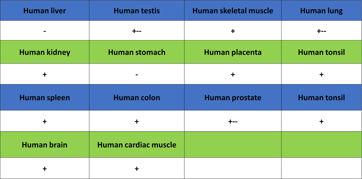
Immunohistochemistry
IHC shows positive staining in paraffin-embedded human cardiac muscle. Anti-Rad51C antibody was used at 1/500 dilution, followed by a HRP Polymer for Rabbit IgG (ready to use). Counterstained with hematoxylin. Heat mediated antigen retrieval with Tris/EDTA buffer pH9.0 was performed before commencing with IHC staining protocol.
IHC shows positive staining in paraffin-embedded human spleen. Anti-Rad51C antibody was used at 1/500 dilution, followed by a HRP Polymer for Rabbit IgG (ready to use). Counterstained with hematoxylin. Heat mediated antigen retrieval with Tris/EDTA buffer pH9.0 was performed before commencing with IHC staining protocol.
IHC shows positive staining in paraffin-embedded human lung cancer. Anti-Rad51C antibody was used at 1/2000 dilution, followed by a HRP Polymer for Rabbit IgG (ready to use). Counterstained with hematoxylin. Heat mediated antigen retrieval with Tris/EDTA buffer pH9.0 was performed before commencing with IHC staining protocol.
IHC shows positive staining in paraffin-embedded human ovarian cancer. Anti-Rad51C antibody was used at 1/500 dilution, followed by a HRP Polymer for Rabbit IgG (ready to use). Counterstained with hematoxylin. Heat mediated antigen retrieval with Tris/EDTA buffer pH9.0 was performed before commencing with IHC staining protocol.
IHC shows positive staining in paraffin-embedded human pancreatic cancer. Anti-Rad51C antibody was used at 1/500 dilution, followed by a HRP Polymer for Rabbit IgG (ready to use). Counterstained with hematoxylin. Heat mediated antigen retrieval with Tris/EDTA buffer pH9.0 was performed before commencing with IHC staining protocol.
IHC shows positive staining in paraffin-embedded human stomach cancer. Anti-Rad51C antibody was used at 1/500 dilution, followed by a HRP Polymer for Rabbit IgG (ready to use). Counterstained with hematoxylin. Heat mediated antigen retrieval with Tris/EDTA buffer pH9.0 was performed before commencing with IHC staining protocol.