Rabbit Anti-Myc Tag Magnetic Agarose is used to immunoprecipitate Myc-tagged fusion proteins from HEK293T cell lysates. During elution with Acidic elution buffer, the enriched Myc-tagged fusion proteins are released from the magnetic agarose.
SDS-PAGE analysis was performed.
Lane 1: Input
Lane 2: Flow-through
Lane 3: Myc-tagged fusion protein
Lane 4: Rabbit IgG Isotype Control Magnetic Agarose
Rabbit Anti-Myc Tag Magnetic Agarose
Rabbit Anti-Myc Tag Magnetic Agarose
Price:
Regular price
$475 USD
Regular price
Sale price
$475 USD
Unit price
per
For shipping services or bulk orders, you may request a quotation.
Secure checkout with

View full details
Product Details
Product Details
Product Specification
| Host | Rabbit |
| Antigen | Myc Tag |
| Immunogen | Synthetic Peptide |
| Antibody Type | Recombinant mAb |
| Isotype | IgG |
| Application | IP, Protein purification, Co-IP, ChIP |
| Purification | Protein A |
| Conjugation | Magnetic Agarose |
| Physical Appearance | Liquid |
| Storage Buffer | 20% slurry in PBS pH7.4, 0.05% Proclin 300 |
| Stability & Storage | 12 months from date of receipt / reconstitution, 2 to 8 °C as supplied |
Background
The c-Myc is a polypeptide protein tag derived from the c-myc gene product, which can be added to the protein by recombinant DNA technology. It contains 10 amino acids (EQKLISEEDL) with a molecular weight of 1.2 KDa and can be fused to the N-terminus or C-terminus of the target protein for purification and detection. The Myc tag is widely used in various expression systems, including bacteria, yeast, insects and mammalian cells, to detect the expression of fusion proteins. It can be used for affinity chromatography and purified by coupling the Myc-tagged antibody to divinylsulfone-activated agarose.
Picture
Picture
SDS-PAGE
Rabbit Anti-Myc Tag Magnetic Agarose is used to immunoprecipitate Myc-tagged fusion proteins from HEK293T cell lysates. During elution with Peptide elution buffer, the enriched Myc-tagged fusion proteins are released from the magnetic agarose.
SDS-PAGE analysis was performed.
Lane 1: Input
Lane 2: Flow-through
Lane 3: Myc-tagged fusion protein
Lane 4: Rabbit IgG Isotype Control Magnetic Agarose
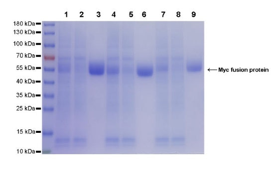
Rabbit Anti-Myc Tag Magnetic Agarose is used to immunoprecipitate different forms of Myc-tagged fusion proteins from HEK293T cell lysates. During elution with Acidic elution buffer, the enriched Myc-tagged fusion proteins are released from the magnetic agarose.
SDS-PAGE analysis was performed.
Lane 1: Input 1
Lane 2: Flow-through 1
Lane 3: Myc-tag at the N-terminus of the fusion protein.
Lane 4: Input 2
Lane 5: Flow-through 2
Lane 6: Myc-tag at the C-terminus of the fusion protein.
Lane 7: Input 3
Lane 8: Flow-through 3
Lane 9: Myc-tag at the internal site of the fusion protein.
ChIP
Chromatin immunoprecipitation (ChIP) was performed on 293F cells were either untransfected (left panel) or transfected with an MYC-tagged human H3 construct (right panel) cross - linked with 1% formaldehyde for 10 min, then chromatin was fragmented by sonication. Parallel reactions used Rabbit Anti-Myc Tag Magnetic Agarose, H3 beads and Rabbit IgG Isotype Control Magnetic Agarose for immunoprecipitation.
Post - immunoprecipitation, both samples were washed, eluted, and cross - links reversed. Purified DNA was analyzed by qPCR.
qPCR (%input: immunoprecipitated DNA/input DNA)
showed the enrichment of RPL30, MYOD1 and SAT-α in
Rabbit Anti-Myc Tag Magnetic Agarose-immunoprecipitated
sample.