WB result of RAB27A Mouse mAb
Primary antibody: TRAB27A Mouse mAb at 1/5000 dilution
Lane 1: HeLa whole cell lysate 20 µg
Lane 2: K562 whole cell lysate 20 µg
Lane 3: Jurkat whole cell lysate 20 µg
Lane 4: HepG2 whole cell lysate 20 µg
Secondary antibody: Goat Anti- mouse IgG, (H+L), HRP conjugated at 1/10000 dilution
Predicted MW: 25 kDa
Observed MW: 26 kDa
RAB27A Mouse mAb (S-M0050)
RAB27A Mouse mAb (S-M0050)
Price:
Regular price
$100 USD
Regular price
Sale price
$100 USD
Unit price
per
For shipping services or bulk orders, you may request a quotation.
Secure checkout with

View full details
Product Details
Product Details
Product Specification
| Host | Mouse |
| Antigen | Rab27A |
| Synonyms | Ras-related protein Rab-27A; RAB27 |
| Location | Lysosome, Endosome |
| Accession | P51159 |
| Clone Number | S-M0050 |
| Antibody Type | Mouse mAb |
| Isotype | IgG1 |
| Application | WB, IHC-P, IF, ICFCM |
| Reactivity | Hu, Ms |
| Positive Sample | HeLa, K562, Jurkat, HepG2, Mouse lung |
| Purification | Protein G |
| Concentration | 1.6 mg/ml |
| Conjugation | Unconjugated |
| Physical Appearance | Liquid |
| Storage Buffer | PBS, 40% Glycerol, 0.05% BSA, 0.02% sodium azide |
| Stability & Storage | 12 months from date of receipt / reconstitution, -20 °C as supplied |
Dilution
| application | dilution | species |
| WB | 1:1000-1:5000 | Hu, Ms |
| IHC-P | 1:5000-1:10000 | Hu, Ms |
| ICFCM | 1:6000 | Hu, Ms |
Background
RAB27A is a small GTPase belonging to the Ras superfamily, encoded by the RAB27A gene, with a molecular weight of approximately 25 kDa. It is primarily localized to secretory vesicles and the plasma membrane within cells, where it functions as a key regulator of vesicular trafficking and exocytosis. RAB27A interacts with various downstream effector proteins in its GTP-bound active form, most notably through binding to synaptotagmin-like proteins (Slps, such as Slp2-a and Slp4-a) and Slac2 family members, thereby regulating vesicle maturation, docking, directed movement, and fusion with the plasma membrane. RAB27A plays important physiological roles in various secretory cell types, particularly in cytotoxic T lymphocytes, where it mediates the polarized secretion of cytolytic granules to enable immune killing functions. In melanocytes, RAB27A forms a ternary complex with myosin Va and melanophilin, which is essential for the transport of melanosomes to dendritic tips. Loss-of-function mutations in the RAB27A gene are the genetic cause of Griscelli syndrome type 2 (GS-2), a disorder characterized by partial albinism (due to impaired melanosome transport) and severe immunodeficiency (due to defective degranulation of cytotoxic T cells and NK cells), often accompanied by hemophagocytic lymphohistiocytosis. Furthermore, aberrant expression of RAB27A is closely associated with tumor invasion and metastasis in various cancers, making it a potential target for research in immunotherapy and tumor metastasis intervention.
Picture
Picture
Western Blot
WB result of RAB27A Mouse mAb
Primary antibody: TRAB27A Mouse mAb at 1/5000 dilution
Lane 1: mouse lung lysate 20 µg
Secondary antibody: Goat Anti- mouse IgG, (H+L), HRP conjugated at 1/10000 dilution
Predicted MW: 25 kDa
Observed MW: 26 kDa
Immunohistochemistry
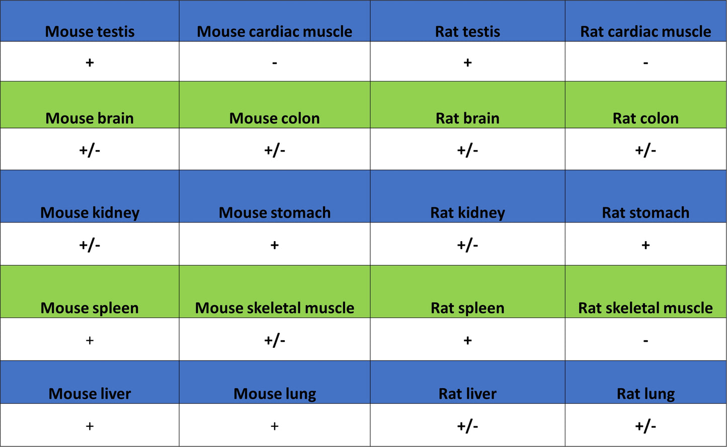
IHC shows positive staining in paraffin-embedded human stomach. Anti-RAB27A antibody was used at 1/10000 dilution, followed by a HRP Polymer for Mouse IgG (ready to use). Counterstained with hematoxylin. Heat mediated antigen retrieval with Tris/EDTA buffer pH9.0 was performed before commencing with IHC staining protocol.
IHC shows positive staining in paraffin-embedded human colon cancer. Anti-RAB27A antibody was used at 1/10000 dilution, followed by a HRP Polymer for Mouse IgG (ready to use). Counterstained with hematoxylin. Heat mediated antigen retrieval with Tris/EDTA buffer pH9.0 was performed before commencing with IHC staining protocol.
IHC shows positive staining in paraffin-embedded mouse stomach. Anti-RAB27A antibody was used at 1/10000 dilution, followed by a HRP Polymer for Mouse IgG (ready to use). Counterstained with hematoxylin. Heat mediated antigen retrieval with Tris/EDTA buffer pH9.0 was performed before commencing with IHC staining protocol.
IHC shows positive staining in paraffin-embedded rat stomach. Anti-RAB27A antibody was used at 1/10000 dilution, followed by a HRP Polymer for Mouse IgG (ready to use). Counterstained with hematoxylin. Heat mediated antigen retrieval with Tris/EDTA buffer pH9.0 was performed before commencing with IHC staining protocol.