WB result of PTPN6 Rabbit pAb
Primary antibody: PTPN6 Rabbit pAb at 1/1000 dilution
Lane 1: Saos-2 whole cell lysate 20 µg
Lane 2: MCF7 whole cell lysate 20 µg
Lane 3: TT whole cell lysate 20 µg
Lane 4: Jurkat whole cell lysate 20 µg
Lane 5: A-431 whole cell lysate 20 µg
Lane 6: K-562 whole cell lysate 20 µg
Lane 7: THP-1 whole cell lysate 20 µg
Negative control: Saos-2 whole cell lysate
Secondary antibody: Goat Anti- rabbit IgG (H+L), HRP conjugated at 1/10000 dilution
Predicted MW: 68 kDa
Observed MW: 68 kDa
PTPN6 Rabbit Polyclonal Antibody
PTPN6 Rabbit Polyclonal Antibody
Price:
Regular price
$100 USD
Regular price
Sale price
$100 USD
Unit price
per
For shipping services or bulk orders, you may request a quotation.
Secure checkout with

View full details
Product Details
Product Details
Product Specification
| Host | Rabbit |
| Antigen | PTPN6 |
| Synonyms | Tyrosine-protein phosphatase non-receptor type 6; Hematopoietic cell protein-tyrosine phosphatase; Protein-tyrosine phosphatase 1C (PTP-1C); Protein-tyrosine phosphatase SHP-1; SH-PTP1; HCP; PTP1C |
| Immunogen | Synthetic Peptide |
| Location | Cytoplasm, Nucleus |
| Accession | P29350 |
| Antibody Type | Polyclonal antibody |
| Isotype | IgG |
| Application | WB, IHC-P |
| Reactivity | Hu, Ms, Rt |
| Positive Sample | MCF7, TT, Jurkat, A-431, K-562, THP-1, RAW264.7, C6, Mouse spleen, mouse thymus, rat spleen, rat thymus, rat liver |
| Purification | Immunogen Affinity |
| Concentration | 0.5 mg/ml |
| Conjugation | Unconjugated |
| Physical Appearance | Liquid |
| Storage Buffer | PBS, 40% Glycerol, 0.05% BSA, 0.02% sodium azide |
| Stability & Storage | 12 months from date of receipt / reconstitution, -20 °C as supplied |
Dilution
| application | dilution | species |
| WB | 1:1000-1:5000 | Hu, Ms, Rt |
| IHC-P | 1:500 | Hu, Ms, Rt |
Background
PTPN6, commonly known as SHP-1 (Src homology region 2 domain-containing phosphatase-1), is a non-receptor protein tyrosine phosphatase predominantly expressed in hematopoietic cells that functions as a critical negative regulator of signal transduction pathways governing immune cell activation, differentiation, and homeostasis. Structurally characterized by two N-terminal Src homology 2 (SH2) domains and a C-terminal catalytic phosphatase domain, PTPN6 exerts its regulatory effects by binding to specific phosphorylated tyrosine residues on activated receptors or adaptor proteins via its SH2 domains, which induces a conformational change that relieves autoinhibition and allows the catalytic domain to dephosphorylate key signaling molecules such as JAK kinases, STAT transcription factors, and receptor tyrosine kinases involved in cytokine and growth factor signaling. Its physiological importance is underscored by the fact that loss-of-function mutations in the PTPN6 gene cause severe autoimmune and inflammatory disorders in both mice (motheaten phenotype) and humans, highlighting its essential role in maintaining immune tolerance and preventing aberrant lymphocyte activation, while its dysregulation has also been implicated in various malignancies and metabolic diseases, making it a significant target for therapeutic intervention in autoimmunity and cancer.
Picture
Picture
Western Blot
WB result of PTPN6 Rabbit pAb
Primary antibody: PTPN6 Rabbit pAb at 1/1000 dilution
Lane 1: RAW 264.7 whole cell lysate 20 µg
Secondary antibody: Goat Anti- rabbit IgG (H+L), HRP conjugated at 1/10000 dilution
Predicted MW: 68 kDa
Observed MW: 68 kDa
WB result of PTPN6 Rabbit pAb
Primary antibody: PTPN6 Rabbit pAb at 1/1000 dilution
Lane 1: mouse marrow lysate 20 µg
Lane 2: mouse spleen lysate 20 µg
Lane 3: mouse thymus lysate 20 µg
Negative control: mouse marrow lysate
Secondary antibody: Goat Anti- rabbit IgG (H+L), HRP conjugated at 1/10000 dilution
Predicted MW: 68 kDa
Observed MW: 68 kDa
WB result of PTPN6 Rabbit pAb
Primary antibody: PTPN6 Rabbit pAb at 1/1000 dilution
Lane 1: C6 whole cell lysate 20 µg
Secondary antibody: Goat Anti- rabbit IgG (H+L), HRP conjugated at 1/10000 dilution
Predicted MW: 68 kDa
Observed MW: 68 kDa
WB result of PTPN6 Rabbit pAb
Primary antibody: PTPN6 Rabbit pAb at 1/1000 dilution
Lane 1: rat brain lysate 20 µg
Lane 2: rat bone marrow lysate 20 µg
Lane 3: rat spleen lysate 20 µg
Lane 4: rat thymus lysate 20 µg
Lane 5: rat liver lysate 20 µg
Negative control: rat brain lysate, rat bone marrow lysate
Secondary antibody: Goat Anti- rabbit IgG (H+L), HRP conjugated at 1/10000 dilution
Predicted MW: 68 kDa
Observed MW: 68 kDa
Immunohistochemistry
IHC shows positive staining in paraffin-embedded human colon. Anti-PTPN6 antibody was used at 1/500 dilution, followed by a HRP Polymer for Rabbit IgG (ready to use). Counterstained with hematoxylin. Heat mediated antigen retrieval with Tris/EDTA buffer pH9.0 was performed before commencing with IHC staining protocol.
IHC shows positive staining in paraffin-embedded human spleen. Anti-PTPN6 antibody was used at 1/500 dilution, followed by a HRP Polymer for Rabbit IgG (ready to use). Counterstained with hematoxylin. Heat mediated antigen retrieval with Tris/EDTA buffer pH9.0 was performed before commencing with IHC staining protocol.
IHC shows positive staining in paraffin-embedded human tonsil. Anti-PTPN6 antibody was used at 1/500 dilution, followed by a HRP Polymer for Rabbit IgG (ready to use). Counterstained with hematoxylin. Heat mediated antigen retrieval with Tris/EDTA buffer pH9.0 was performed before commencing with IHC staining protocol.
IHC shows positive staining in paraffin-embedded human lung cancer. Anti-PTPN6 antibody was used at 1/500 dilution, followed by a HRP Polymer for Rabbit IgG (ready to use). Counterstained with hematoxylin. Heat mediated antigen retrieval with Tris/EDTA buffer pH9.0 was performed before commencing with IHC staining protocol.
IHC shows positive staining in paraffin-embedded human ovarian cancer. Anti-PTPN6 antibody was used at 1/500 dilution, followed by a HRP Polymer for Rabbit IgG (ready to use). Counterstained with hematoxylin. Heat mediated antigen retrieval with Tris/EDTA buffer pH9.0 was performed before commencing with IHC staining protocol.
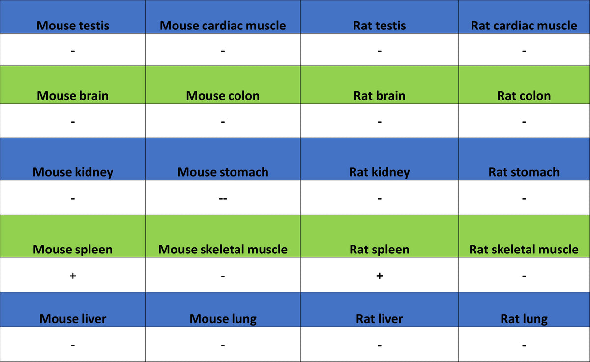
IHC shows positive staining in paraffin-embedded mouse spleen. Anti-PTPN6 antibody was used at 1/500 dilution, followed by a HRP Polymer for Rabbit IgG (ready to use). Counterstained with hematoxylin. Heat mediated antigen retrieval with Tris/EDTA buffer pH9.0 was performed before commencing with IHC staining protocol.
IHC shows positive staining in paraffin-embedded rat spleen. Anti-PTPN6 antibody was used at 1/500 dilution, followed by a HRP Polymer for Rabbit IgG (ready to use). Counterstained with hematoxylin. Heat mediated antigen retrieval with Tris/EDTA buffer pH9.0 was performed before commencing with IHC staining protocol.