WB result of PSMB9 Recombinant Rabbit mAb
Primary antibody: PSMB9 Recombinant Rabbit mAb at 1/1000 dilution
Lane 1: THP-1 whole cell lysate 20 μg
Lane 2: Ramos whole cell lysate 20 μg
Lane 3: Raji whole cell lysate 20 μg
Lane 4: Daudi whole cell lysate 20 μg
Secondary antibody: Goat Anti-Rabbit IgG (H+L), HRP conjugated at 1/10000 dilution
Predicted MW: 23 kDa
Observed MW: 21, 23 kDa
PSMB9 Recombinant Rabbit mAb (S-4183-122)
PSMB9 Recombinant Rabbit mAb (S-4183-122)
Price:
Regular price
$100 USD
Regular price
Sale price
$100 USD
Unit price
per
For shipping services or bulk orders, you may request a quotation.
Secure checkout with

View full details
Product Details
Product Details
Product Specification
| Host | Rabbit |
| Antigen | PSMB9 |
| Immunogen | Recombinant Protein |
| Location | Cytoplasm, Nucleus |
| Accession | P28065 |
| Clone Number | S-4183-122 |
| Antibody Type | Recombinant mAb |
| Isotype | IgG |
| Application | WB, IHC-P |
| Reactivity | Hu, Ms, Rt |
| Positive Sample | THP-1, Ramos, Raji, Daudi, A20, mouse spleen, PC-12, rat spleen, rat liver |
| Purification | Protein A |
| Concentration | 0.5 mg/ml |
| Conjugation | Unconjugated |
| Physical Appearance | Liquid |
| Storage Buffer | PBS, 40% Glycerol, 0.05% BSA, 0.02% sodium azide |
| Stability & Storage | 12 months from date of receipt / reconstitution, -20 °C as supplied |
Dilution
| application | dilution | species |
| WB | 1:1000-1:2000 | Hu, Ms, Rt |
| IHC-P | 1:1000 | Hu |
Background
PSMB9 (proteasome subunit beta type 9) is a catalytic subunit of the immunoproteasome encoded by the PSMB9 gene located in the MHC class II region on human chromosome 6, also known as β1i or LMP2, containing approximately 219 amino acids with a molecular weight of about 23 kDa. As a core component of the immunoproteasome, PSMB9 replaces the PSMB6 subunit of the constitutive proteasome under interferon-γ induction, thereby altering the cleavage preference of the proteasome and enhancing its ability to cleave peptides after hydrophobic and basic residues, which is crucial for generating antigenic peptides suitable for binding to MHC class I molecules. Functionally, PSMB9 participates in antigen processing and presentation, and its expression is broadly correlated with T cell infiltration in the tumor microenvironment, expression of immune checkpoint genes, and activation of interferon response pathways, with high expression generally predicting better responses to immune checkpoint inhibitors and CAR-T cell therapy. In addition, PSMB9 may exert tumor-suppressive effects in melanoma, where its low expression is associated with poor prognosis, while vemurafenib can induce ferroptosis by activating PSMB9 to inhibit melanoma progression. Clinically, variants such as G156D in PSMB9 can cause proteasome-associated autoinflammatory syndrome, manifesting as immunodeficiency and autoinflammation.
Picture
Picture
Western Blot
WB result of PSMB9 Recombinant Rabbit mAb
Primary antibody: PSMB9 Recombinant Rabbit mAb at 1/1000 dilution
Lane 1: untreated HeLa whole cell lysate 20 μg
Lane 2: HeLa treated with 100ng/mL hIFN-γ for 48 hours whole cell lysate 20 μg
Secondary antibody: Goat Anti-Rabbit IgG (H+L), HRP conjugated at 1/10000 dilution
Predicted MW: 23 kDa
Observed MW: 21 kDa
WB result of PSMB9 Recombinant Rabbit mAb
Primary antibody: PSMB9 Recombinant Rabbit mAb at 1/1000 dilution
Lane 1: A20 whole cell lysate 20 μg
Lane 2: mouse spleen lysate 20 μg
Secondary antibody: Goat Anti-Rabbit IgG (H+L), HRP conjugated at 1/10000 dilution
Predicted MW: 23 kDa
Observed MW: 21 kDa
WB result of PSMB9 Recombinant Rabbit mAb
Primary antibody: PSMB9 Recombinant Rabbit mAb at 1/1000 dilution
Lane 1: PC-12 whole cell lysate 20 μg
Lane 2: rat spleen lysate 20 μg
Lane 3: rat liver lysate 20 μg
Secondary antibody: Goat Anti-Rabbit IgG (H+L), HRP conjugated at 1/10000 dilution
Predicted MW: 23 kDa
Observed MW: 21, 23 kDa
Immunohistochemistry
IHC shows positive staining in paraffin-embedded human brain. Anti-PSMB9 antibody was used at 1/1000 dilution, followed by a HRP Polymer for Rabbit IgG (ready to use). Counterstained with hematoxylin. Heat mediated antigen retrieval with Tris/EDTA buffer pH9.0 was performed before commencing with IHC staining protocol.
IHC shows positive staining in paraffin-embedded human prostate. Anti-PSMB9 antibody was used at 1/1000 dilution, followed by a HRP Polymer for Rabbit IgG (ready to use). Counterstained with hematoxylin. Heat mediated antigen retrieval with Tris/EDTA buffer pH9.0 was performed before commencing with IHC staining protocol.
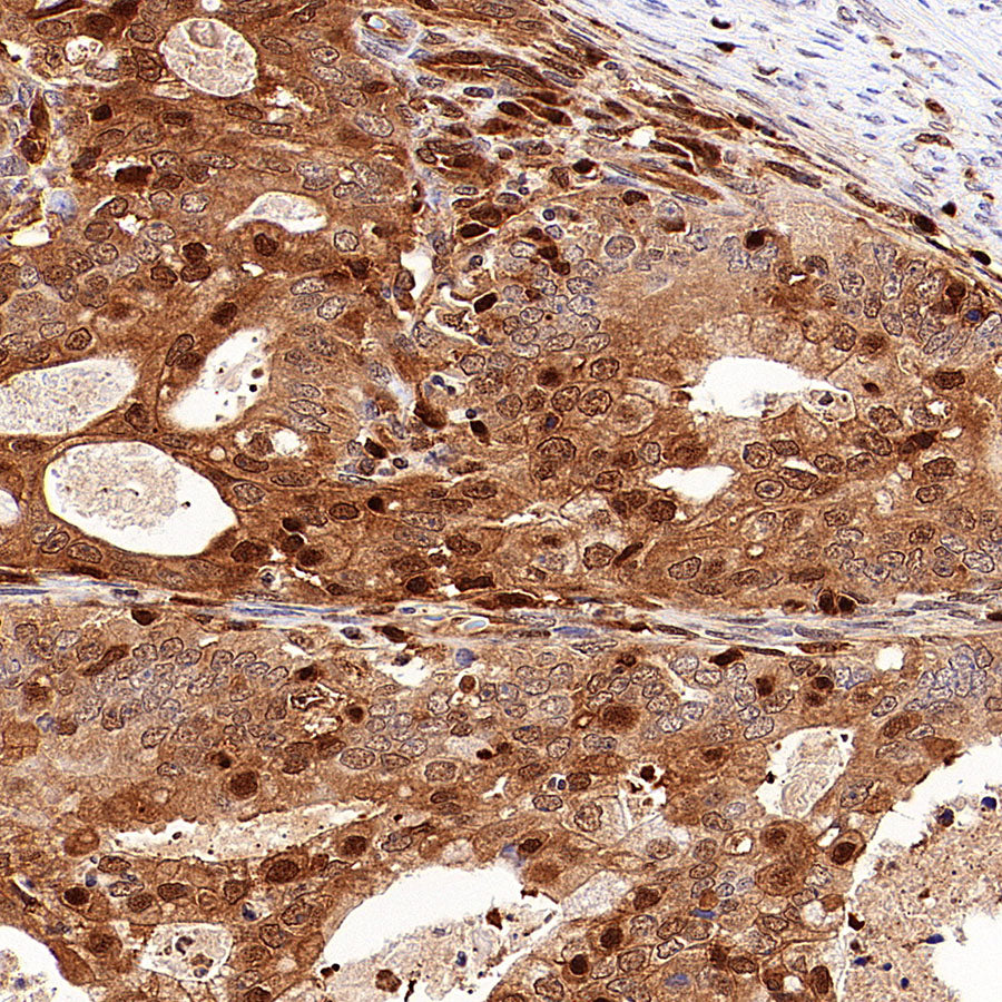
IHC shows positive staining in paraffin-embedded human endometrial cancer. Anti-PSMB9 antibody was used at 1/1000 dilution, followed by a HRP Polymer for Rabbit IgG (ready to use). Counterstained with hematoxylin. Heat mediated antigen retrieval with Tris/EDTA buffer pH9.0 was performed before commencing with IHC staining protocol.