WB result of Phosphotyrosine Rabbit mAb
Primary antibody: Phosphotyrosine Rabbit mAb at 1/1000 dilution
Lane 1: HeLa whole cell lysate 20 µg
Lane 2: HeLa treated with Sodium pervanadate (3 mM, 30 min) whole cell lysate 20 µg
Secondary antibody: Goat Anti-Rabbit IgG, (H+L), HRP conjugated at 1/10000 dilution
Predicted MW: multiple
Observed MW: multiple
Exposure time: 180 s
Phosphotyrosine Rabbit mAb (S-R207)
Phosphotyrosine Rabbit mAb (S-R207)
Price:
Regular price
$110 USD
Regular price
Sale price
$110 USD
Unit price
per
For shipping services or bulk orders, you may request a quotation.
Secure checkout with

View full details
Product Details
Product Details
Product Specification
| Host | Rabbit |
| Antigen | Phospho-Tyrosine |
| Synonyms | p‑Tyr, pY, tyrosine phosphorylation |
| Clone Number | S-R207 |
| Antibody Type | Recombinant mAb |
| Isotype | IgG |
| Application | WB, IHC-P, IP |
| Reactivity | Hu, Ms, Rt |
| Purification | Protein A |
| Concentration | 0.5 mg/ml |
| Conjugation | Unconjugated |
| Physical Appearance | Liquid |
| Storage Buffer | PBS, 40% Glycerol, 0.05%BSA, 0.03% Proclin 300 |
| Stability & Storage | 12 months from date of receipt / reconstitution, -20 °C as supplied |
Dilution
| application | dilution | species |
| Dot Blot | 1:500 | |
| WB | 1:1000 | |
| IP | 1:50 | |
| IHC | 1:500 |
Background
Tyrosine phosphorylation is the addition of a phosphate (PO43−) group to the amino acid tyrosine on a protein. It is one of the main types of protein phosphorylation. This transfer is made possible through enzymes called tyrosine kinases. Tyrosine phosphorylation is a key step in signal transduction and the regulation of enzymatic activity.
Picture
Picture
Western Blot
IP
Phosphotyrosine Rabbit mAb at 1/50 dilution (1 µg) immunoprecipitating Phospho-Tyrosine in 0.4 mg HeLa treated with Sodium pervanadate (3 mM, 30 min) whole cell lysate.
Western blot was performed on the immunoprecipitate using Phospho-Tyrosine Rabbit mAb at 1/1000 dilution.
Secondary antibody (HRP) for IP was used at 1/400 dilution.
Lane 1: HeLa treated with Sodium pervanadate (3mM, 30min) whole cell lysate 20 µg (Input)
Lane 2: Phospho-Tyrosine Rabbit mAb IP in HeLa treated with Sodium pervanadate (3mM, 30min) whole cell lysate
Lane 3: Rabbit monoclonal IgG IP in HeLa treated with Sodium pervanadate (3mM, 30min) whole cell lysate
Predicted MW: multiple
Observed MW: multiple
Exposure time: 30 s
Dot Blot
Dot blot result of Phosphotyrosine Rabbit mAb
Lane 1: Modified peptide library (Phospho-Serine)
Lane 2: Modified peptide library (Phospho-Threonine)
Lane 3: Modified peptide library (Phospho-Tyrosine)
Lane 4: Unmodified peptide library (Serine)
Lane 5: Unmodified peptide library (Threonine)
Lane 6: Unmodified peptide library (Tyrosine)
Primary antibody: Phosphotyrosine Rabbit mAb at 1/500 dilution
Secondary antibody: Goat Anti-rabbit IgG, (H+L), HRP conjugated at 1/10000 dilution
Immunohistochemistry
IHC shows positive staining in paraffin-embedded mouse lung (left) and negative staining in phosphatase treated (37 °C, 2h) paraffin-embedded mouse lung (right). Anti- Phospho-Tyrosine antibody was used at 1/500 dilution, followed by a HRP Polymer for Mouse & Rabbit IgG (ready to use). Counterstained with hematoxylin. Heat mediated antigen retrieval with Tris/EDTA buffer pH9.0 was performed before commencing with IHC staining protocol.
IHC shows positive staining in paraffin-embedded rat lung (left) and negative staining in phosphatase treated (37 °C, 2h) paraffin-embedded mouse lung (right). Anti- Phospho-Tyrosine antibody was used at 1/500 dilution, followed by a HRP Polymer for Mouse & Rabbit IgG (ready to use). Counterstained with hematoxylin. Heat mediated antigen retrieval with Tris/EDTA buffer pH9.0 was performed before commencing with IHC staining protocol.
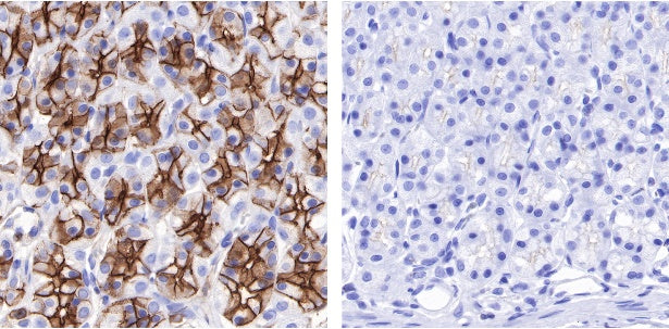
IHC shows positive staining in paraffin-embedded rat stomach (left) and negative staining in phosphatase treated (37 °C, 2h) paraffin-embedded mouse lung (right). Anti- Phospho-Tyrosine antibody was used at 1/500 dilution, followed by a HRP Polymer for Mouse & Rabbit IgG (ready to use). Counterstained with hematoxylin. Heat mediated antigen retrieval with Tris/EDTA buffer pH9.0 was performed before commencing with IHC staining protocol.