WB result of Phosphotyrosine Recombinant Mouse mAb
Primary antibody: Phosphotyrosine Recombinant Mouse mAb at 1/1000 dilution
Lane 1: untreated A431 whole cell lysate 20 µg
Lane 2: A431 treated with 50 mM Pervanadate for 5 minutes whole cell lysate 20 µg
Secondary antibody: Goat Anti-mouse IgG, (H+L), HRP conjugated at 1/10000 dilution
Predicted MW: Multiple
Observed MW: Multiple
Phosphotyrosine Mouse mAb (S-R433-1)
Phosphotyrosine Mouse mAb (S-R433-1)
Price:
Regular price
$110 USD
Regular price
Sale price
$110 USD
Unit price
per
For shipping services or bulk orders, you may request a quotation.
Secure checkout with

View full details
Product Details
Product Details
Product Specification
| Host | Mouse |
| Synonyms | p‑Tyr, pY, tyrosine phosphorylation |
| Clone Number | S-R433-1 |
| Antibody Type | Mouse mAb |
| Isotype | IgG2b,k |
| Application | WB, ICC |
| Reactivity | Hu, Ms |
| Purification | Protein A |
| Concentration | 2 mg/ml |
| Conjugation | Unconjugated |
| Physical Appearance | Liquid |
| Storage Buffer | PBS, 40% Glycerol, 0.05% BSA, 0.03% Proclin 300 |
| Stability & Storage | 12 months from date of receipt / reconstitution, -20 °C as supplied |
Dilution
| application | dilution | species |
| Dot Blot | 1:2000 | |
| WB | 1:1000 | |
| ICC | 1:500 |
Background
Tyrosine phosphorylation refers to the addition of a phosphate group to the hydroxyl oxygen of the tyrosine amino acid residue in a protein. This process is catalyzed by specific kinases, such as protein tyrosine kinases (PTKs), which transfer the gamma phosphate group of ATP to the tyrosine hydroxyl group. Tyrosine phosphorylation is involved in the regulation of protein function, activity, and localization. It plays a key role in signal transduction pathways, mediating cellular responses to extracellular stimuli.
Picture
Picture
Western Blot
WB result of Phosphotyrosine Recombinant Mouse mAb
Primary antibody: Phosphotyrosine Recombinant Mouse mAb at 1/1000 dilution
Lane 1: untreated NIH/3T3 whole cell lysate 20 µg
Lane 2: NIH/3T3 treated with 10 mM Pervanadate for 20 minutes whole cell lysate 20 µg
Secondary antibody: Goat Anti-mouse IgG, (H+L), HRP conjugated at 1/10000 dilution
Predicted MW: Multiple
Observed MW: Multiple
Dot Blot
Dot blot result of Phosphotyrosine Mouse mAb
Lane 1: Modified peptide library (Phospho-Serine)
Lane 2: Modified peptide library (Phospho-Threonine)
Lane 3: Modified peptide library (Phospho-Tyrosine)
Lane 4: Unmodified peptide library (Serine)
Lane 5: Unmodified peptide library (Threonine)
Lane 6: Unmodified peptide library (Tyrosine)
Primary antibody: Phosphotyrosine Mouse mAb at 1/2000 dilution
Secondary antibody: Goat Anti-mouse IgG, (H+L), HRP conjugated at 1/10000 dilution
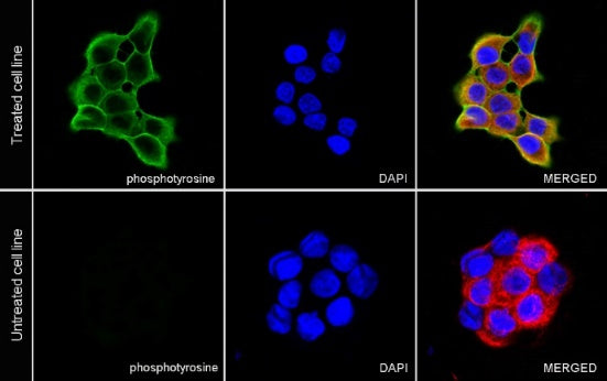
Immunocytochemistry
ICC analysis of A431 cells treated with Pervanadate (50mM, 5min) (top panel) and A431 cells untreated with Pervanadate (50mM, 5min) (below panel). Anti- Phosphotyrosine antibody was used at 1/500 dilution (Green) and incubated overnight at 4°C. Goat polyclonal Antibody to Mouse IgG - H&L (Alexa Fluor® 488) was used as secondary antibody at 1/1000 dilution. The cells were fixed with 100% ice-cold methanol and permeabilized with 0.1% PBS-Triton X-100. Nuclei were counterstained with DAPI (Blue). Counterstain with tubulin (Red).