WB result of Phosphothreonine Recombinant Rabbit mAb
Primary antibody: Phosphothreonine Recombinant Rabbit mAb at 1/1000 dilution
Lane 1: untreated HeLa whole cell lysate 20 µg
Lane 2: HeLa treated with 100ng/ml Calyculin A for 30mintes whole cell lysate 20 µg
Secondary antibody: Goat Anti-Rabbit IgG (H+L), HRP conjugated at 1/10000 dilution
Predicted MW: Multiple
Observed MW: Multiple
Phosphothreonine Rabbit mAb (S-4181-29)
Phosphothreonine Rabbit mAb (S-4181-29)
Price:
Regular price
$135 USD
Regular price
Sale price
$135 USD
Unit price
per
For shipping services or bulk orders, you may request a quotation.
Secure checkout with

View full details
Product Details
Product Details
Product Specification
| Host | Rabbit |
| Antigen | Phosphothreonine |
| Synonyms | Phosphothreonine, threonine phosphorylation, p-Thr, pT |
| Immunogen | Synthetic Peptide |
| Clone Number | S-4181-29 |
| Antibody Type | Recombinant mAb |
| Isotype | IgG |
| Application | WB, ICC |
| Purification | Protein A |
| Concentration | 1 mg/ml |
| Conjugation | Unconjugated |
| Physical Appearance | Liquid |
| Storage Buffer | PBS, 40% Glycerol, 0.05% BSA, 0.02% sodium azide |
| Stability & Storage | 12 months from date of receipt / reconstitution, -20 °C as supplied |
Dilution
| application | dilution | species |
| Dot Blot | 1:1000 | Species Independent |
| WB | 1:1000 | Species Independent |
| ICC | 1:100 | Species Independent |
Background
Phosphothreonine (pThr) is the phosphorylated ester of threonine and an important phosphoamino acid. It serves as a key participant in reversible protein phosphorylation—a core cellular signaling mechanism—and plays vital roles in both eukaryotes and prokaryotes. Similar to phosphorylation modification, its function is primarily achieved through two means: first, as a molecular "switch" that induces conformational changes in the modified protein to alter its activity; and second, as a "Lego block" that serves as a recognition site for specific structural domains (such as FHA domains), thereby mediating protein–protein interactions and assembling signaling complexes. Interestingly, structural studies have shown that phosphothreonine induces a more pronounced disorder-to-order transition in proteins compared to phosphoserine, and preferentially adopts a compact cyclic conformation, making its conformational changes more like a "step function" rather than the "rheostat"-like fine-tuning induced by phosphoserine. In living organisms, free phosphothreonine is scarce, but as an important post-translational modification form of proteins, it remains a core component in the regulatory networks governing numerous life processes.
Picture
Picture
Western Blot
WB result of Phosphothreonine Recombinant Rabbit mAb
Primary antibody: Phosphothreonine Recombinant Rabbit mAb at 1/1000 dilution
Lane 1: untreated NIH/3T3 whole cell lysate 20 µg
Lane 2: NIH/3T3 treated with 100ng/ml Calyculin A for 30mintes whole cell lysate 20 µg
Secondary antibody: Goat Anti-Rabbit IgG (H+L), HRP conjugated at 1/10000 dilution
Predicted MW: Multiple
Observed MW: Multiple
WB result of Phosphothreonine Recombinant Rabbit mAb
Primary antibody: Phosphothreonine Recombinant Rabbit mAb at 1/1000 dilution
Lane 1: untreated C6 whole cell lysate 20 µg
Lane 2: C6 treated with 100ng/ml Calyculin A for 30mintes whole cell lysate 20 µg
Secondary antibody: Goat Anti-Rabbit IgG (H+L), HRP conjugated at 1/10000 dilution
Predicted MW: Multiple
Observed MW: Multiple
Dot Blot
Dot blot result of Phosphothreonine Recombinant Rabbit mAb
Lane 1: Modified peptide library (Phospho-Serine)
Lane 2: Modified peptide library (Phospho-Threonine)
Lane 3: Modified peptide library (Phospho-Tyrosine)
Lane 4: Unmodified peptide library (Serine)
Lane 5: Unmodified peptide library (Threonine)
Lane 6: Unmodified peptide library (Tyrosine)
Primary antibody: Phosphothreonine Recombinant Rabbit mAb at 1/1000 dilution
Secondary antibody: Goat Anti-rabbit IgG, (H+L), HRP conjugated at 1/10000 dilution
Immunocytochemistry
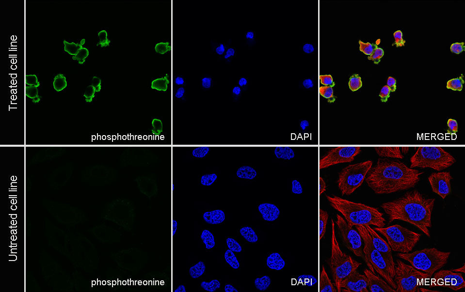
ICC analysis of HeLa cells treated with Calyculin A (100nM/ml, 30min) (top panel) and untreated HeLa cells (below panel). Anti-Phosphothreonine antibody was used at 1/100 dilution (Green) and incubated overnight at 4°C. Goat polyclonal Antibody to Rabbit IgG - H&L (Alexa Fluor® 488) was used as secondary antibody at 1/1000 dilution. The cells were fixed with 4% PFA and permeabilized with 0.1% PBS-Triton X-100. Nuclei were counterstained with DAPI (Blue). Counterstain with tubulin (Red).