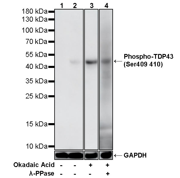
WB result of Phospho-TDP43 (Ser409/410) Recombinant Rabbit mAb
Primary antibody: Phospho-TDP43 (Ser409/410) Recombinant Rabbit mAb at 1/1000 dilution
Lane 1: 293F transfected with empty vector whole cell lysate 20 µg
Lane 2: 293F transfected with wild-type TDP43-His fusion protein whole cell lysate 20 µg
Lane 3: 293F transfected with wild-type TDP43-His fusion protein then treated with 1uM Okadaic Acid for 60 minutes whole cell lysate 20 µg
Lane 4: 293F transfected with wild-type TDP43-His fusion protein then treated with 1uM Okadaic Acid for 60 minutes whole cell lysate 20 µg (phosphatase-treated membrane)
Secondary antibody: Goat Anti-rabbit IgG (H+L), HRP conjugated at 1/10000 dilution
Predicted MW: 45 kDa
Observed MW: 45 kDa
This blot was developed with high sensitivity substrate
Phospho-TDP43 (Ser409/410) Recombinant Rabbit mAb (S-3862)
Phospho-TDP43 (Ser409/410) Recombinant Rabbit mAb (S-3862)

Product Details
Product Details
Product Specification
| Host | Rabbit |
| Antigen | Phospho-TDP43 (Ser409/410) |
| Synonyms | TAR DNA-binding protein 43; TDP43; TARDBP |
| Location | Cytoplasm, Nucleus, Mitochondrion |
| Accession | Q13148 |
| Clone Number | S-3862 |
| Antibody Type | Recombinant mAb |
| Isotype | IgG |
| Application | WB |
| Reactivity | Hu |
| Purification | Protein A |
| Concentration | 0.5 mg/ml |
| Conjugation | Unconjugated |
| Physical Appearance | Liquid |
| Storage Buffer | PBS, 40% Glycerol, 0.05% BSA, 0.02% sodium azide |
| Stability & Storage | 12 months from date of receipt / reconstitution, -20 °C as supplied |
Dilution
| application | dilution | species |
| WB | 1:1000 | Hu |
Background
Phospho-TDP43 (Ser409/410) refers to the abnormally phosphorylated form of the TAR DNA-binding protein 43 (TDP-43) at serine residues 409 and 410, which is widely recognized as the pathological hallmark and biochemical signature of TDP-43 proteinopathies, including amyotrophic lateral sclerosis (ALS) and frontotemporal lobar degeneration (FTLD). Under physiological conditions, TDP-43 is predominantly nuclear and exists in a non-phosphorylated state; however, in disease, it becomes hyperphosphorylated at these C-terminal serine residues, leading to its mislocalization to the cytoplasm and incorporation into ubiquitin-positive inclusions. Studies using monoclonal antibodies specifically generated against the dual-phosphorylated Ser409/410 epitope (mAb pS409/410) have demonstrated that this modification is a highly consistent and specific feature of pathological TDP-43 across the entire spectrum of sporadic and familial TDP-43 proteinopathies, while normal nuclear TDP-43 remains unstained. Notably, phosphorylation at both Ser409 and Ser410 is required for antibody recognition, and casein kinase 1 (CK1) has been identified as a candidate kinase responsible for generating this pathological epitope. Functionally, this phosphorylation event promotes TDP-43 oligomerization and enhances its fibrillation propensity in vitro, contributing to the formation of the characteristic sarkosyl-insoluble, urea-soluble aggregates found in affected neurons and glial cells. Additionally, the phosphorylation status at these sites serves as a critical biomarker for the neuropathological diagnosis of TDP-43 proteinopathies and is increasingly recognized as a potential therapeutic target, with recent studies identifying CDC7 as another kinase capable of phosphorylating these residues and driving neurotoxicity in disease models.
Picture
Picture
Western Blot