WB result of Phospho-SRC (Tyr419) Recombinant Rabbit mAb
Primary antibody: Phospho-SRC (Tyr419) Recombinant Rabbit mAb at 1/1000 dilution
Lane 1: untreated A431 whole cell lysate 20 µg
Lane 2: A431 treated with 50 mM pervanadate for 5 minutes whole cell lysate 20 µg
Secondary antibody: Goat Anti-rabbit IgG, (H+L), HRP conjugated at 1/10000 dilution
Predicted MW: 60 kDa
Observed MW: 60 kDa
Phospho-SRC (Tyr419) Recombinant Rabbit mAb (S-1182-5)
Phospho-SRC (Tyr419) Recombinant Rabbit mAb (S-1182-5)
Price:
Regular price
$100 USD
Regular price
Sale price
$100 USD
Unit price
per
For shipping services or bulk orders, you may request a quotation.
Secure checkout with

View full details
Product Details
Product Details
Product Specification
| Host | Rabbit |
| Antigen | Phospho-SRC (Tyr419) |
| Synonyms | Proto-oncogene tyrosine-protein kinase Src, Proto-oncogene c-Src, pp60c-src (p60-Src) |
| Immunogen | Synthetic Peptide |
| Location | Cytoplasm, Nucleus, Cell membrane |
| Accession | P12931 |
| Clone Number | S-1182-5 |
| Antibody Type | Recombinant mAb |
| Isotype | IgG |
| Application | WB, ICC, IP |
| Reactivity | Hu, Rt |
| Predicted Reactivity | Zf, Fs, Rt, Ms, Av, Dg, Xe, Bv, Pg |
| Purification | Protein A |
| Concentration | 0.5 mg/ml |
| Conjugation | Unconjugated |
| Physical Appearance | Liquid |
| Storage Buffer | PBS, 40% Glycerol, 0.05% BSA, 0.03% Proclin 300 |
| Stability & Storage | 12 months from date of receipt / reconstitution, -20 °C as supplied |
Dilution
| application | dilution | species |
| Dot Blot | 1:1000 | |
| WB | 1:1000 | Hu, Rt |
| IP | 1:50 | Hu |
| ICC | 1:100 | Hu |
Background
Proto-oncogene tyrosine-protein kinase Src, also known as proto-oncogene c-Src, or simply c-Src belongs to a family of Src family kinases. c-Src phosphorylates specific tyrosine residues in other tyrosine kinases. It plays a role in the regulation of embryonic development and cell growth. An elevated level of activity of c-Src is suggested to be linked to cancer progression by promoting other signals. Mutations in c-Src could be involved in the malignant progression of colon cancer. The activation of c-Src causes the dephosphorylation of the tyrosine 527. This induces long-range allostery via protein domain dynamics, causing the structure to be destabilized, resulting in the opening up of the SH3, SH2 and kinase domains and the autophosphorylation of the residue tyrosine 419.
Picture
Picture
Western Blot
WB result of Phospho-SRC (Tyr419) Recombinant Rabbit mAb
Primary antibody: Phospho-SRC (Tyr419) Recombinant Rabbit mAb at 1/1000 dilution
Lane 1: Untreated C6 whole cell lysate 20 µg
Lane 2: C6 treated with pervanadate(10mM, 20min) whole cell lysate 20 µg
Negative control: Untreated C6 whole cell lysate 20 µg
Secondary antibody: Goat Anti-Rabbit IgG, (H+L), HRP conjugated at 1/10000 dilution Predicted MW: 60 kDa
Observed MW: 60kDa
IP
Phospho-SRC (Tyr419) Recombinan Rabbit mAb at 1/50 dilution (1 µg) immunoprecipitating Phospho-SRC (Tyr419) in 0.4 mg A431 treated with 50mM pervanadate for 5 minutes whole cell lysate.
Western blot was performed on the immunoprecipitate using Phospho-SRC (Tyr419) Recombinan Rabbit mAb at 1/1000 dilution.
Secondary antibody (HRP) for IP was used at 1/1000 dilution.
Lane 1: A431 treated with 50mM pervanadate for 5 minutes whole cell lysate 20 µg (Input)
Lane 2: Phospho-SRC (Tyr419) Rabbit mAb IP in A431 treated with 50mM pervanadate for 5 minutes whole cell lysate
Lane 3: Rabbit monoclonal IgG IP in A431 treated with 50mM pervanadate for 5 minutes whole cell lysate
Predicted MW: 60 kDa
Observed MW: 60kDa
Dot Blot
Dot blot result of Phospho-SRC (Tyr419) Recombinant Rabbit mAb
Lane 1: Phospho-SRC (Tyr419) peptide
Lane 2: SRC unmodified peptide
Primary antibody: Phospho-SRC (Tyr419) Recombinant Rabbit mAb at 1/1000 dilution
Secondary antibody: Goat Anti-rabbit IgG, (H+L), HRP conjugated at 1/10000 dilution
Immunocytochemistry
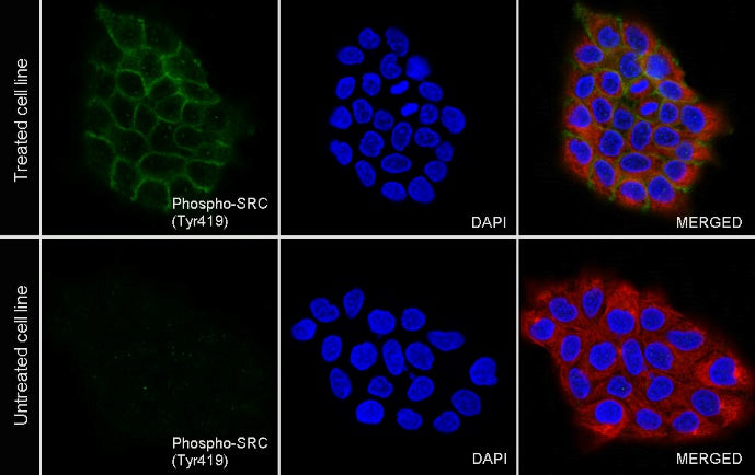
ICC analysis of A431 cells treated with 50mM pervanadate for 5 minutes (top panel) and untreated A431 cells (below panel). Anti- Phospho-SRC (Tyr419) antibody was used at 1/100 dilution (Green) and incubated overnight at 4°C. Goat polyclonal Antibody to Rabbit IgG - H&L (Alexa Fluor® 488) was used as secondary antibody at 1/1000 dilution. The cells were fixed with 100% ice-cold methanol and permeabilized with 0.1% PBS-Triton X-100. Nuclei were counterstained with DAPI (Blue). Counterstain with tubulin (Red).