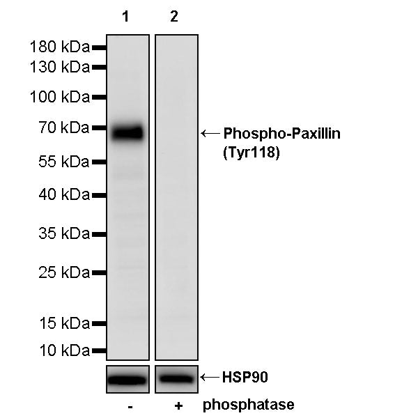
WB result of Phospho-Paxillin (Tyr118) Recombinant Rabbit mAb
Blocking/Diluting buffer and concentration: 5% NFDM/TBST
Primary antibody incubation conditions: overnight at 4°C
Primary antibody: Phospho-Paxillin (Tyr118) Recombinant Rabbit mAb at 1/1000 dilution
Lane 1: C2C12 whole cell lysate 20 µg
Lane 2: C2C12 whole cell lysate 20 µg (phosphatase treated membrane)
Secondary antibody: Goat Anti- rabbit IgG, (H+L), HRP conjugated at 1/10000 dilution
Predicted MW: 64 kDa
Observed MW: 68 kDa
This blot was developed with high sensitivity substrate
Phospho-Paxillin (Tyr118) Recombinant Rabbit mAb (S-4990)
Phospho-Paxillin (Tyr118) Recombinant Rabbit mAb (S-4990)
Price:
Regular price
$100 USD
Regular price
Sale price
$100 USD
Unit price
per
For shipping services or bulk orders, you may request a quotation.
Secure checkout with

View full details
Product Details
Product Details
Product Specification
| Host | Rabbit |
| Antigen | Phospho-Paxillin (Tyr118) |
| Synonyms | Paxillin; PXN |
| Location | Cytoplasm, Cytoskeleton |
| Accession | P49023 |
| Clone Number | S-4990 |
| Antibody Type | Recombinant mAb |
| Isotype | IgG |
| Application | WB |
| Reactivity | Hu, Ms, Rt, Mk |
| Positive Sample | C2C12, C6 |
| Purification | Protein A |
| Concentration | 0.5 mg/ml |
| Conjugation | Unconjugated |
| Physical Appearance | Liquid |
| Storage Buffer | PBS, 40% Glycerol, 0.05% BSA, 0.02% sodium azide |
| Stability & Storage | 12 months from date of receipt / reconstitution, -20 °C as supplied |
Dilution
| application | dilution | species |
| WB | 1:500-1:1000 | Hu, Ms, Rt, Mk |
Background
Phospho-Paxillin (Tyr118) refers to the phosphorylated form of paxillin at tyrosine residue 118, serving as a key activation marker of cell adhesion, cytoskeletal reorganization, and signal transduction. Paxillin is a multi-domain adaptor protein localized to focal adhesions, and its Tyr118 site can be phosphorylated by non-receptor tyrosine kinases such as FAK (focal adhesion kinase) and Src family kinases. This phosphorylation event provides a high-affinity binding site for SH2 domain-containing effector proteins (e.g., Crk, Dock180), subsequently activating small GTPases such as Rac1 and Cdc42 to regulate cell migration, invasion, and survival. Following cell adhesion to the extracellular matrix, integrin-mediated FAK activation rapidly upregulates p-Paxillin (Tyr118) levels, promoting focal adhesion dynamics and protrusion formation; conversely, its phosphorylation level decreases significantly during cell detachment or under stress conditions. This modification is particularly critical in tumor invasion and metastasis, as high expression of p-Paxillin (Tyr118) is closely associated with the metastatic potential and poor prognosis of various solid tumors, including breast cancer and colorectal cancer. Additionally, p-Paxillin (Tyr118) participates in physiological processes such as neuronal axon guidance, regulation of cardiomyocyte contractile function, and wound healing, and is often used as a biomarker to assess cell migration activity and evaluate drug efficacy targeting the focal adhesion kinase signaling pathway.
Picture
Picture
Western Blot
WB result of Phospho-Paxillin (Tyr118) Recombinant Rabbit mAb
Blocking/Diluting buffer and concentration: 5% NFDM/TBST
Primary antibody incubation conditions: overnight at 4°C
Primary antibody: Phospho-Paxillin (Tyr118) Recombinant Rabbit mAb at 1/1000 dilution
Lane 1: C6 whole cell lysate 20 µg
Lane 2: C6 whole cell lysate 20 µg (phosphatase treated membrane)
Secondary antibody: Goat Anti- rabbit IgG, (H+L), HRP conjugated at 1/10000 dilution
Predicted MW: 64 kDa
Observed MW: 68 kDa