WB result of Phospho-p44/42 MAPK (Erk1/2) (Thr202/Tyr204) Recombinant Rabbit mAb
Blocking/Diluting buffer and concentration: 5% NFDM/TBST
Primary antibody incubation conditions: overnight at 4°C
Primary antibody: Phospho-p44/42 MAPK (Erk1/2) (Thr202/Tyr204) Recombinant Rabbit mAb at 1/1000 dilution
Lane 1: untreated HeLa whole cell lysate 20 µg
Lane 2: HeLa starve overnight, then treated with 200 nM TPA for 4 hours whole cell lysate 20 µg
Secondary antibody: Goat Anti- rabbit IgG, (H+L), HRP conjugated at 1/10000 dilution
Predicted MW: 41, 43 kDa
Observed MW: 39 kDa
This blot was developed with high sensitivity substrate
Phospho-p44/42 MAPK (Erk1/2) (Thr202/Tyr204) Recombinant Rabbit mAb (S-5119)
Phospho-p44/42 MAPK (Erk1/2) (Thr202/Tyr204) Recombinant Rabbit mAb (S-5119)
Price:
Regular price
$100 USD
Regular price
Sale price
$100 USD
Unit price
per
For shipping services or bulk orders, you may request a quotation.
Secure checkout with

View full details
Product Details
Product Details
Product Specification
| Host | Rabbit |
| Antigen | Phospho-p44/42 MAPK (Erk1/2) (Thr202/Tyr204) |
| Synonyms | Mitogen-activated protein kinase 1; MAP kinase 1; MAPK 1; ERT1; Extracellular signal-regulated kinase 2 (ERK-2); MAP kinase isoform p42 (p42-MAPK); Mitogen-activated protein kinase 2 (MAP kinase 2; MAPK 2); ERK2; PRKM1; PRKM2; MAPK1 |
| Location | Cytoplasm, Cytoskeleton, Nucleus |
| Accession | P28482、P27361 |
| Clone Number | S-5119 |
| Antibody Type | Recombinant mAb |
| Isotype | IgG |
| Application | WB, IHC-P |
| Reactivity | Hu, Ms, Rt |
| Purification | Protein A |
| Concentration | 0.5 mg/ml |
| Conjugation | Unconjugated |
| Physical Appearance | Liquid |
| Storage Buffer | PBS, 40% Glycerol, 0.05% BSA, 0.02% sodium azide |
| Stability & Storage | 12 months from date of receipt / reconstitution, -20 °C as supplied |
Dilution
| application | dilution | species |
| WB | 1:500-1:1000 | Hu, Ms, Rt |
| IHC-P | 1:500 | Hu, Ms, Rt |
Background
Phospho-p44/42 MAPK (Erk1/2) (Thr202/Tyr204) is the activated form of extracellular signal-regulated kinases 1 and 2 (ERK1/2) following phosphorylation at specific sites, specifically referring to the simultaneous phosphorylation of threonine and tyrosine residues at positions 202/204 of ERK1 (corresponding to positions 183/185 of ERK2, using the classic dual-site designation). This phosphorylation event is specifically catalyzed by the upstream kinases MEK1/2 and serves as a core hallmark of activation of the canonical Ras-Raf-MEK-ERK signaling pathway. Once activated by phosphorylation, p-ERK1/2 translocates to the nucleus, where it phosphorylates and regulates various downstream substrates, including transcription factors (such as Elk-1, c-Fos, and c-Myc) and cell cycle regulatory proteins, thereby participating in the regulation of key cell fate decisions including proliferation, differentiation, survival, migration, and apoptosis. This phosphorylation modification is tightly regulated and, under normal physiological conditions, responds to extracellular stimuli such as growth factors, cytokines, and mitogens. In various pathological states, particularly in more than one-third of malignant tumors (such as melanoma, colorectal cancer, and pancreatic cancer), the phosphorylation level at these sites is often persistently and abnormally elevated due to upstream activating mutations (e.g., RAS or BRAF mutations), driving uncontrolled tumor cell proliferation and metastasis. Therefore, detecting the level of Phospho-p44/42 MAPK (Thr202/Tyr204) not only serves to assess the activation status of the ERK signaling pathway but also acts as a key biomarker for screening the efficacy of MEK/ERK-targeted inhibitors and monitoring resistance mechanisms.
Picture
Picture
Western Blot
WB result of Phospho-p44/42 MAPK (Erk1/2) (Thr202/Tyr204) Recombinant Rabbit mAb
Blocking/Diluting buffer and concentration: 5% NFDM/TBST
Primary antibody incubation conditions: overnight at 4°C
Primary antibody: Phospho-p44/42 MAPK (Erk1/2) (Thr202/Tyr204) Recombinant Rabbit mAb at 1/1000 dilution
Lane 1: untreated NIH/3T3 whole cell lysate 20 µg
Lane 2: NIH/3T3 starve overnight, then treated with 100 ng/ml PDGF for 5 minutes whole cell lysate 20 µg
Secondary antibody: Goat Anti- rabbit IgG, (H+L), HRP conjugated at 1/10000 dilution
Predicted MW: 41, 43 kDa
Observed MW: 39, 42 kDa
This blot was developed with high sensitivity substrate
Immunohistochemistry
IHC shows positive staining in paraffin-embedded human kidney. Anti- Phospho-p44/42 MAPK (Erk1/2) (Thr202/Tyr204) antibody was used at 1/500 dilution, followed by a HRP Polymer for Rabbit IgG (ready to use). Counterstained with hematoxylin. Heat mediated antigen retrieval with Tris/EDTA buffer pH9.0 was performed before commencing with IHC staining protocol.
IHC shows positive staining in paraffin-embedded human spleen. Anti- Phospho-p44/42 MAPK (Erk1/2) (Thr202/Tyr204) antibody was used at 1/500 dilution, followed by a HRP Polymer for Rabbit IgG (ready to use). Counterstained with hematoxylin. Heat mediated antigen retrieval with Tris/EDTA buffer pH9.0 was performed before commencing with IHC staining protocol.
IHC shows positive staining in paraffin-embedded human colon cancer. Anti- Phospho-p44/42 MAPK (Erk1/2) (Thr202/Tyr204) antibody was used at 1/500 dilution, followed by a HRP Polymer for Rabbit IgG (ready to use). Counterstained with hematoxylin. Heat mediated antigen retrieval with Tris/EDTA buffer pH9.0 was performed before commencing with IHC staining protocol.
IHC shows positive staining in paraffin-embedded human ovarian cancer. Anti- Phospho-p44/42 MAPK (Erk1/2) (Thr202/Tyr204) antibody was used at 1/500 dilution, followed by a HRP Polymer for Rabbit IgG (ready to use). Counterstained with hematoxylin. Heat mediated antigen retrieval with Tris/EDTA buffer pH9.0 was performed before commencing with IHC staining protocol.
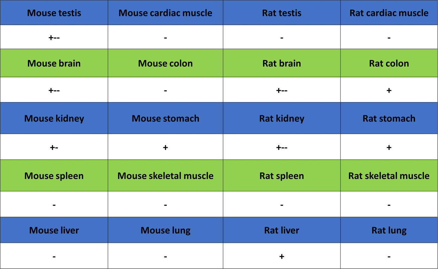
IHC shows positive staining in paraffin-embedded mouse stomach. Anti- Phospho-p44/42 MAPK (Erk1/2) (Thr202/Tyr204) antibody was used at 1/500 dilution, followed by a HRP Polymer for Rabbit IgG (ready to use). Counterstained with hematoxylin. Heat mediated antigen retrieval with Tris/EDTA buffer pH9.0 was performed before commencing with IHC staining protocol.
IHC shows positive staining in paraffin-embedded rat colon. Anti- Phospho-p44/42 MAPK (Erk1/2) (Thr202/Tyr204) antibody was used at 1/500 dilution, followed by a HRP Polymer for Rabbit IgG (ready to use). Counterstained with hematoxylin. Heat mediated antigen retrieval with Tris/EDTA buffer pH9.0 was performed before commencing with IHC staining protocol.