WB result of Phospho-NF-κB p65 (S529) Recombinant Rabbit mAb
Primary antibody: Phospho-NF-κB p65 (S529) Recombinant Rabbit mAb at 1/1000 dilution
Lane 1: untreated HeLa whole cell lysate 20 µg
Lane 2: HeLa treated with 20 ng/ml TNF-alpha for 5 miniutes whole cell lysate 20 µg
Secondary antibody: Goat Anti-Rabbit IgG, (H+L), HRP conjugated at 1/10000 dilution Predicted MW: 60 kDa
Observed MW: 65 kDa
Phospho-NF-κB p65 (S529) Recombinant Rabbit mAb (S-1125-59)
Phospho-NF-κB p65 (S529) Recombinant Rabbit mAb (S-1125-59)
Price:
Regular price
$100 USD
Regular price
Sale price
$100 USD
Unit price
per
For shipping services or bulk orders, you may request a quotation.
Secure checkout with

View full details
Product Details
Product Details
Product Specification
| Host | Rabbit |
| Antigen | Phospho-NF-κB p65 (S529) |
| Synonyms | Transcription factor p65; Nuclear factor NF-kappa-B p65 subunit; Nuclear factor of kappa light polypeptide gene enhancer in B-cells 3; RELA; NFKB3 |
| Immunogen | Synthetic Peptide |
| Location | Cytoplasm, Nucleus |
| Clone Number | S-1125-59 |
| Antibody Type | Recombinant mAb |
| Isotype | IgG |
| Application | WB, ChIP |
| Reactivity | Hu |
| Positive Sample | HeLa |
| Purification | Protein A |
| Concentration | 0.5 mg/ml |
| Conjugation | Unconjugated |
| Physical Appearance | Liquid |
| Storage Buffer | PBS, 40% Glycerol, 0.05% BSA, 0.03% Proclin 300 |
| Stability & Storage | 12 months from date of receipt / reconstitution, -20 °C as supplied |
Dilution
| application | dilution | species |
| Dot Blot | 1:1000 | |
| WB | 1:1000 | Hu |
| ChIP | 1:20-1:50 | Hu |
Background
Phosphorylation of NF-κB p65 at Ser529 is a post-translational modification that plays a significant role in the regulation of NF-κB activity. This modification is catalyzed by casein kinase II (CK2) upon stimulation by cytokines such as interleukin-1β (IL-1β) or tumor necrosis factor α (TNFα), leading to increased p65 transactivation potential in a gene-specific manner. Phosphorylation at S529 enhances the transcriptional activity of NF-κB, which is crucial for regulating various cellular processes including inflammation, immune responses, and cell survival. Dysregulation of NF-κB signaling, including phosphorylation at S529, has been implicated in various diseases, particularly in the context of chronic inflammation and aging. Phosphorylation of p65 at S529 has also been reported to promote tumorigenesis in various tissues, including the bone, breast, and head and neck, suggesting a significant role of NF-κB phosphorylation in different types of cancers.
Picture
Picture
Western Blot
Dot Blot
Dot blot result of Phospho-NF-κB p65 (S529) Recombinant Rabbit mAb
Lane 1: NF-κB p65 (S529) phospho peptide
Lane 2: NF-κB p65 unmodified peptide
Primary antibody: Phospho-NF-κB p65 (S529) Recombinant Rabbit mAb at 1/1000 dilution
Secondary antibody: Goat Anti-rabbit IgG, (H+L), HRP conjugated at 1/10000 dilution
ChIP
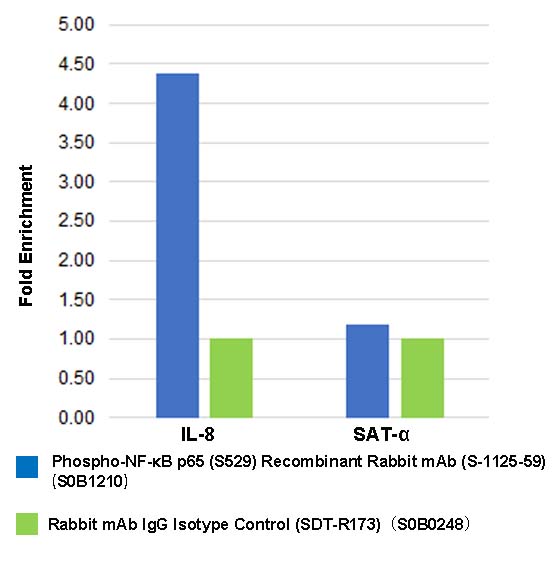
Chromatin immunoprecipitation (ChIP) was performed on HeLa+hTNF-α (20 ng/ml, 5 min) cells cross-linked with 1% formaldehyde for 10 min, then chromatin was fragmented by sonication.
Parallel reactions used Phospho NF-κB p65 (S529) Recombinant Rabbit mAb (S-1125-59) and Rabbit mAb IgG Isotype Control (SDT-R173) at 1:50 for immunoprecipitation. Post - immunoprecipitation, both samples were washed, eluted, and cross-links reversed. Purified DNA was analyzed by qPCR.
qPCR showed the enrichment of IL-8 and SAT-α in Phospho-NF-κB
p65 (S529) Recombinant Rabbit mAb (S-1125-59)-immunoprecipitated sample.