WB result of Phospho-ERK1 (Thr202) + ERK2 (Thr185) Recombinant Rabbit mAb
Primary antibody: Phospho-ERK1 (Thr202) + ERK2 (Thr185) Recombinant Rabbit mAb at 1/1000 dilution
Lane 1: untreated Jurkat whole cell lysate 20 μg
Lane 2: Jurkat treated with 200ng/mL PMA for 30 min whole cell lysate 20 μg
Secondary antibody: Goat Anti-Rabbit IgG (H+L), HRP conjugated at 1/10000 dilution
Predicted MW: 42 kDa
Observed MW: 39 kDa
Phospho-ERK1 (Thr202) + ERK2 (Thr185) Recombinant Rabbit mAb (S-6265)
Phospho-ERK1 (Thr202) + ERK2 (Thr185) Recombinant Rabbit mAb (S-6265)
Price:
Regular price
$100 USD
Regular price
Sale price
$100 USD
Unit price
per
For shipping services or bulk orders, you may request a quotation.
Secure checkout with

View full details
Product Details
Product Details
Product Specification
| Host | Rabbit |
| Antigen | Phospho-ERK1 (Thr202) + ERK2 (Thr185) |
| Synonyms | Mitogen-activated protein kinase 3; MAP kinase 3; MAPK 3; ERT2; Extracellular signal-regulated kinase 1 (ERK-1); Insulin-stimulated MAP2 kinase; MAP kinase isoform p44 (p44-MAPK); Microtubule-associated protein 2 kinase; p44-ERK1; ERK1; PRKM3; MAPK3 |
| Location | Cytoplasm, Nucleus |
| Accession | P27361、P28482 |
| Clone Number | S-6265 |
| Antibody Type | Recombinant mAb |
| Isotype | IgG |
| Application | WB, IHC-P |
| Reactivity | Hu, Ms, Rt |
| Purification | Protein A |
| Concentration | 0.5 mg/ml |
| Conjugation | Unconjugated |
| Physical Appearance | Liquid |
| Storage Buffer | PBS, 40% Glycerol, 0.05% BSA, 0.02% sodium azide |
| Stability & Storage | 12 months from date of receipt / reconstitution, -20 °C as supplied |
Dilution
| application | dilution | species |
| WB | 1:1000 | Hu, Ms, Rt |
| IHC | 1:100 | Hu |
Background
Phospho-ERK1 (Thr202) and Phospho-ERK2 (Thr185) refer, respectively, to the active forms of extracellular signal-regulated kinase 1 (ERK1, p44 MAPK) and ERK2 (p42 MAPK) phosphorylated at the threonine residue within the TEY motif of their activation loop (Thr202 of ERK1 and Thr185 of ERK2). ERK1/2 are the terminal effector kinases of the Ras–Raf–MEK–ERK signaling cascade; MEK1/2 first catalyzes phosphorylation of Tyr204/187 and then phosphorylation of Thr202/185, and dual phosphorylation at these two sites is required for full enzyme activation. This phosphorylation event enables ERK1/2 to translocate into the nucleus and phosphorylate transcription factors such as Elk-1 and c-Fos, thereby regulating diverse processes including cell proliferation, survival, differentiation, migration, and metabolism. Notably, detection of phosphorylation at the Thr202/185 sites alone generally reflects the activation status of ERK1/2, but phosphorylation at this site per se is not sufficient to fully represent kinase activity, because nuclear translocation of ERK and effective binding to substrates are also subject to multiple layers of regulation, including dimerization, scaffold proteins, and docking sites. In approximately one-third of human cancers, Ras–Raf–MEK–ERK pathway activity is elevated; therefore, phosphorylated ERK1/2 serves both as a commonly used marker of activation of this pathway and as a key pharmacodynamic indicator that must be monitored in anticancer drug development.
Picture
Picture
Western Blot
WB result of Phospho-ERK1 (Thr202) + ERK2 (Thr185) Recombinant Rabbit mAb
Primary antibody: Phospho-ERK1 (Thr202) + ERK2 (Thr185) Recombinant Rabbit mAb at 1/1000 dilution
Lane 1: untreated PC-12 whole cell lysate 20 μg
Lane 2: PC-12 serum starvation for 4 hours, then treated with 100ng/mL β-NGF for 5 min whole cell lysate 20 μg
Secondary antibody: Goat Anti-Rabbit IgG (H+L), HRP conjugated at 1/10000 dilution
Predicted MW: 42 kDa
Observed MW: 42 kDa
Immunohistochemistry
IHC shows positive staining in paraffin-embedded human ovarian cancer. Anti-Phospho-ERK1(Thr202)+ERK2(Thr185) antibody was used at 1/100 dilution, followed by a HRP Polymer for Rabbit IgG (ready to use). Counterstained with hematoxylin. Heat mediated antigen retrieval with Tris/EDTA buffer pH9.0 was performed before commencing with IHC staining protocol.
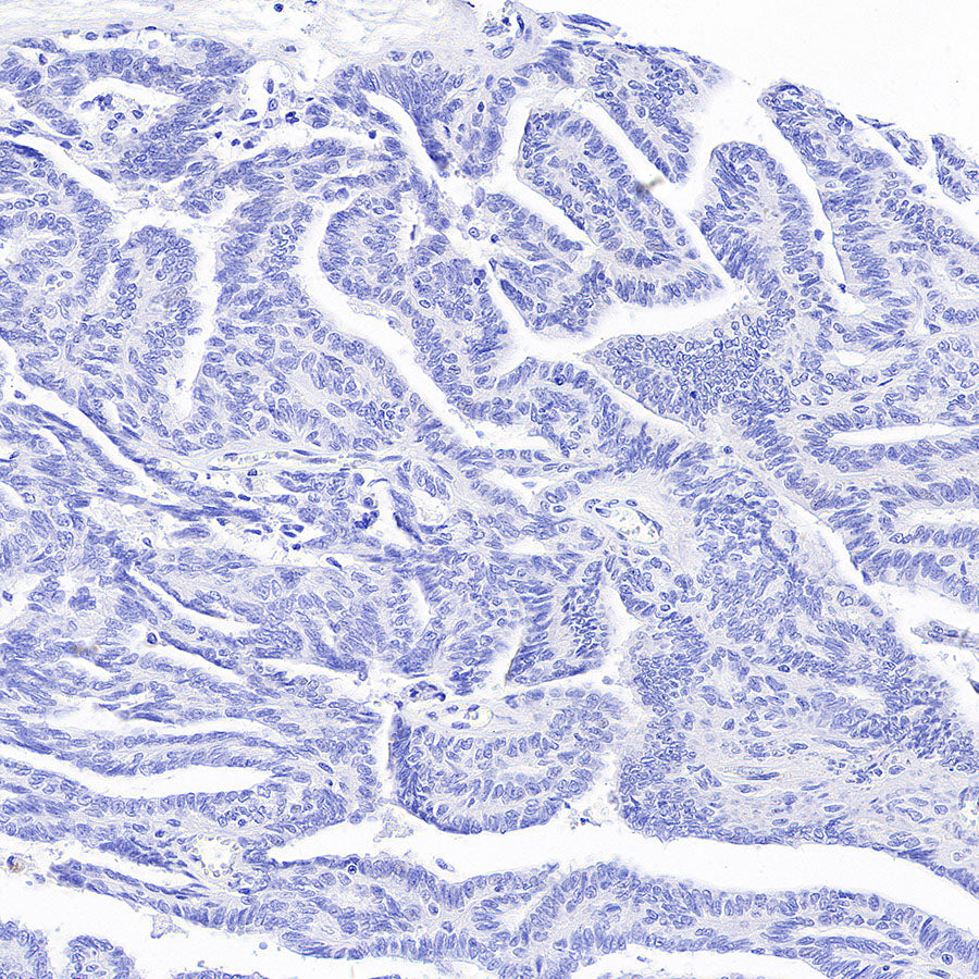
Negative control: IHC shows negative staining in paraffin-embedded human ovarian cancer treated with phosphatase. Anti-Phospho-ERK1(Thr202)+ERK2(Thr185) antibody was used at 1/100 dilution, followed by a HRP Polymer for Rabbit IgG (ready to use). Counterstained with hematoxylin. Heat mediated antigen retrieval with Tris/EDTA buffer pH9.0 was performed before commencing with IHC staining protocol.